
Recombinant Proteins
A recombinant protein is produced by cloning the gene for a target protein into an expression vector and expressing it in a host cell line, rather than purifying the native protein from tissue. The host determines what happens to the protein after translation: mammalian cells such as HEK293 and CHO add human-like glycosylation and support the natural folding of complex extracellular domains, while E. coli produces non-glycosylated protein at lower cost, often requiring refolding from inclusion bodies. Tag position and conjugation state, His, Fc or site-specific Avi-tag biotin, then determine how a protein can be captured, immobilised or detected in a given assay.
Browse Recombinant ProteinsRecombinant Protein Categories
Recombinant proteins in our range are organised by biological role and application, from soluble cytokines used in cell culture to membrane receptor ectodomains used in binding and capture assays. Choose a category below to browse that part of the range.
Active EGFR and Biotinylated PD-L1: Two Functionally Validated Proteins
EGFR and PD-L1 anchor two of the most studied pathways in cancer research: receptor tyrosine kinase signalling and T cell checkpoint inhibition. Both proteins below are supplied with functional assay data confirming ligand or antibody binding, not purity data alone.

Human EGFR Protein (Active Recombinant, C-His) (STJP000982)
Produced in HEK293 cells spanning the Met1-Ser645 region with a C-terminal His tag, and less than 0.1 EU/µg endotoxin by LAL method. Binding activity is confirmed by functional ELISA, where immobilised EGFR captures its ligand EGF across a 7-25 ng/mL linear range. EGFR is a receptor tyrosine kinase that activates the RAS-RAF-MEK-ERK and PI3K-AKT cascades on ligand binding, and is a well-established driver in several solid tumour types.1
View product →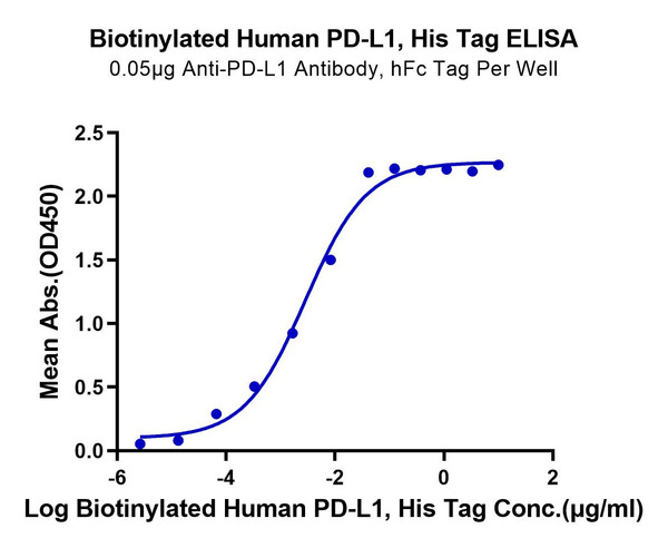
Human PD-L1/B7-H1 Protein (C-His-Avi, Biotin) (STJP001643)
Site-specifically biotinylated through the Avi-tag/BirA system for defined-orientation capture, with purity above 95% by SEC-HPLC and a dose-response EC50 of 2.8 ng/mL against an anti-PD-L1 antibody by ELISA. PD-L1 engagement of PD-1 delivers an inhibitory signal that limits T cell activation and proliferation, a pathway central to checkpoint blockade research.2
View product →Cytokines and Growth Factors
Cytokines are small secreted proteins that regulate cell signalling in the immune system and beyond, binding specific receptors to trigger transduction pathways that control gene expression. Interleukin-2 (IL-2) drives T cell proliferation and, together with its receptor subunits CD25 (IL-2RA) and CD122 (IL-2RB), is used to study T cell growth signalling and regulatory T cell homeostasis.3 IL-4, IL-6 and IL-7 cover Th2 differentiation, acute-phase and B cell responses, and T cell survival signalling respectively, GM-CSF drives monocyte to dendritic cell differentiation, and type I interferon IFN-alpha 2 supports antiviral and innate immune signalling studies.
| Target | Notes | Products |
|---|---|---|
| IL-2 | T cell growth factor; ED50 <0.1 ng/mL by CTLL-2 proliferation assay | |
| IL-2RA/CD25 | IL-2 receptor alpha chain, marks activated and regulatory T cells | |
| IL-2RB/CD122 | Shared subunit of the high and low affinity IL-2 receptor complexes | STJP000431 |
| IL-4 | Drives Th2 differentiation and IgE class switching | STJP000226 |
| IL-6 | Acute-phase response and B cell differentiation cytokine | STJP000183 |
| IL-7 | Supports T cell survival and homeostatic proliferation | STJP000231 |
| GM-CSF (CSF2) | Drives monocyte to dendritic cell differentiation, often paired with IL-4 | |
| IFN-alpha 2 | Type I interferon involved in antiviral and innate immune signalling | STJP001424 |
Immune Checkpoint and Costimulatory Proteins
PD-1 and its ligand PD-L1 form the checkpoint axis exploited by tumours to blunt cytotoxic T cell activity.2 CTLA-4 acts further upstream in T cell priming, competing with CD28 for the CD80/CD86 ligands on antigen-presenting cells and functioning as an essential negative regulator of T cell responses.5 CD28 itself delivers the costimulatory signal required alongside T cell receptor engagement for full T cell activation.5
| Target | Notes | Products |
|---|---|---|
| PD-1/PDCD1/CD279 | Dose-response EC50 of 0.19 µg/mL against PD-L1 by ELISA on the biotinylated format | |
| PD-L1/B7-H1/CD274 | See Featured Products above for the biotinylated format with ELISA validation data | |
| CTLA-4/CD152 | Dose-response EC50 of 15.4 ng/mL by ELISA on the biotinylated format | |
| CD28 | Dose-response EC50 of 24.4 ng/mL by ELISA on the biotinylated, multi-species format | |
Cancer and Receptor Tyrosine Kinase Signalling
EGFR (ErbB1/HER1) and HER2 (ErbB2) belong to the same four-member receptor tyrosine kinase family and frequently dimerise together in tumour cells, with the EGFR-HER2 heterodimer among the most robust signalling pairs in the family despite HER2 having no known ligand of its own.1 EGFRvIII is a constitutively active deletion variant of EGFR that removes part of the ligand-binding domain and recurs in glioblastoma research models.
| Target | Notes | Products |
|---|---|---|
| EGFR/ErbB1/HER1 | Active format confirmed by EGF binding ELISA; see Featured Products above | |
| HER2/ErbB2/CD340 | Active recombinant formats available alongside biotinylated capture reagents | |
| EGFRvIII | Constitutively active deletion variant, biotinylated via Avi-tag; EC50 of 16.1 ng/mL by ELISA | STJP001704 |
Vascular and Angiogenesis Research
VEGFR-2 (KDR/CD309) is the principal receptor mediating VEGF-driven endothelial cell proliferation, migration and new vessel formation, and is regulated through a layered system of receptor-interacting proteins, endocytosis and intracellular trafficking.4
| Target | Notes | Products |
|---|---|---|
| VEGFR-2/KDR/CD309 | Dose-response EC50 of 0.35 µg/mL by ELISA on the biotinylated format | |
Viral Antigens
Viral antigens in our range are produced from defined strain or variant sequences for use in serology, ELISA coating and cross-reactivity studies, with lot-to-lot consistency confirmed on the product datasheet. Antigens from different strains or variants can differ substantially in their antigenic, functional and structural properties, so tag position and strain should be checked against the assay before ordering. The range covers SARS-CoV-2 and its variants, MERS, HIV, hepatitis B and C, cytomegalovirus and H5N1 avian influenza.
| Antigen | Notes | Products |
|---|---|---|
| SARS-CoV-2 Spike | S1, full-length trimer and Omicron (B.1.1.529) RBD formats; S trimer confirmed to bind ACE2-Fc across a 23 ng/mL linear range by ELISA | |
| SARS-CoV-2 Nucleocapsid | His-tagged, wild-type and Omicron variant formats | STJP000259 |
| MERS coronavirus | Spike S1, S2 and RBD, plus nucleocapsid, for serology and cross-reactivity work against SARS-CoV-2 | |
| HIV-1 envelope glycoproteins | gp120 and gp160 envelope glycoproteins used in serology and envelope binding studies | |
| Hepatitis B surface antigen (Adw2) | Produced in P. pastoris yeast; validated for ELISA and western blot | STJP000337 |
| Hepatitis C Core protein | Structural core protein for serology panels | STJP000248 |
| Influenza A H5N1 Hemagglutinin | Produced in insect cells; Indonesia and Vietnam clade strains | STJP000616 |
| Cytomegalovirus pp52 | Tegument phosphoprotein used in CMV serology | STJP001885 |
Stem Cell and Organoid Factors
Embryonic and induced pluripotent stem cells self-renew and can differentiate into lineages from all three germ layers, and their growth and differentiation in culture are directed by recombinant growth factors and cytokines that mimic the natural signals cells encounter in the body. Organoids, which are derived from stem cells, offer a more physiologically relevant alternative to 2D culture for preclinical work, and depend on the same class of recombinant factors to regulate self-organisation, proliferation and differentiation. Haematopoietic stem cells are directed down the lymphoid lineage, producing T cells, B cells and NK cells, or the myeloid lineage, producing neutrophils, macrophages and dendritic cells, according to the cytokine and growth factor signals they receive.
| Factor | Notes | Products |
|---|---|---|
| TGF-β1 / Activin A | Pluripotency maintenance and early lineage specification cues | STJP000387 |
| Noggin (NOG) | Used in brain, intestine and pancreas organoid protocols, often paired with Wnt | STJP001239 |
| Wnt3a | Intestinal, liver and retina organoid media; check carrier-free formats for media making | STJP004984 |
| BMP4 | Liver and kidney organoid protocols, typically used as a time-limited pulse for patterning | STJP004044 |
Tags and Conjugation Options
Recombinant proteins in our range are supplied unconjugated or conjugated to a range of tags, depending on the assay. Affinity tags such as 6xHis, GST and FLAG support purification and capture, while Fc fusions to human IgG1 improve stability and can increase avidity through dimerisation. Site-specific biotinylation through the Avi-tag/BirA system labels a single defined lysine residue, giving a consistent biotin to protein ratio for streptavidin-based capture. Tags are positioned at either the N- or C-terminus of the protein depending on the target and are stated on the product datasheet.
| Type | Examples | Use case |
|---|---|---|
| Affinity tags | 6xHis, GST, FLAG, Strep-tag | Purification and capture; ELISA coating; pull-downs |
| Fusion partners | Fc (IgG1), albumin | Improve stability, support dimerisation and increase avidity |
| Site-specific biotin | Avi-tag (BirA) | Defined biotin stoichiometry; streptavidin capture |
| Direct labels | Biotin, HRP, fluorescent dyes | Direct detection; BLI, ELISA and flow standards |
Expression Systems
The expression host determines the post-translational modifications a protein carries, which in turn affects folding, activity and how directly the result translates to a native human target. Choose a host that matches the downstream application.
| System | PTMs | Strengths | Considerations |
|---|---|---|---|
| HEK293 (human) | Human-like glycosylation | Best for receptors and Fc fusions; native folding | Higher cost than bacterial systems |
| CHO (mammalian) | Mammalian glycosylation | Robust secretion; consistent lot-to-lot performance | Longer production timelines |
| Insect (Sf9/Sf21) | Limited glycosylation | High yields for secreted extracellular domains | Glycan patterns differ from human |
| E. coli | No glycosylation | Scalable and cost-effective | May require refolding; endotoxin control needed |
| Yeast (Pichia) | High-mannose glycans | Good secreted yields; simple media | Non-human glycans can affect binding for some targets |
Quality and QC Attributes
Every recombinant protein datasheet states the expression system, tag position, purity method and endotoxin result for that specific lot, so the specifications below describe what the majority of the range is produced to rather than a guarantee for every individual product.
| Attribute | Typical spec / info | Why it matters |
|---|---|---|
| Purity | ≥95% by SDS-PAGE or SEC-HPLC | Minimises off-target effects and assay interference |
| Endotoxin | ≤1 EU/µg (≤0.1 ng/µg) by LAL gel clot method | Critical for cell culture and immune assays |
| Bioactivity | Cell-based EC50, or ligand/receptor binding by ELISA, SPR or BLI | Confirms functional performance beyond purity alone |
| Sequence & tag | UniProt reference, signal peptide, His/Fc/GST or Avi-biotin | Assay compatibility and capture or immobilisation orientation |
| Formulation | Carrier-free, or with BSA/trehalose | Affects suitability for conjugation and certain assays |
| Storage | Aliquot at -80°C; avoid 3 or more freeze-thaw cycles | Preserves activity across experiments |
Formulation and Storage
Match formulation to the assay: carrier-free protein suits conjugation and biophysical work, while a stabiliser suits long-term storage or low-abundance dosing.
| Formulation | Pros | Considerations |
|---|---|---|
| Carrier-free (no BSA) | Ideal for conjugation and biophysics | Use low-protein-bind tubes; add trehalose if storing |
| With carrier (BSA, trehalose) | Improves stability against surface adsorption | Not suitable for some conjugations or assays |
| Lyophilised | Room-temperature shipping; long shelf life | Follow reconstitution instructions carefully |
| Liquid (frozen) | Ready to use | Aliquot on receipt; avoid repeat freeze-thaw |
Reconstitution and Dosing
Use sterile PBS or water, with carrier protein where recommended on the datasheet, and spin vials briefly before opening. The volumes below are typical starting points.
| Vial content | Target concentration | Add this volume | Notes |
|---|---|---|---|
| 10 µg | 0.1 mg/mL | 100 µL diluent | Gives 100 µL at 100 µg/mL |
| 50 µg | 0.5 mg/mL | 100 µL diluent | Gives 100 µL at 0.5 mg/mL |
| 100 µg | 0.2 mg/mL | 500 µL diluent | Aliquot for single use |
| 1 mg | 1 mg/mL | 1 mL diluent | Prepare working stocks as needed |
Assay Standards and Controls
Controls improve quantitation and troubleshooting across ELISA, BLI/SPR, neutralisation and cell-based assays.
| Type | Where used | Notes |
|---|---|---|
| Calibrators/standards | ELISA and qPCR protein calibration | Define the dynamic range and limit of detection |
| Capture antigens | ELISA plates, serology | Choose tag or biotin format for orientation control |
| Ligand/binding pairs | BLI/SPR kinetics | Avi-biotin plus streptavidin sensor surface |
| Neutralisation controls | Cell-based assays | Active cytokine paired with a blocking antibody |
Protein FAQs
| Question | Answer |
|---|---|
| Carrier-free or with carrier, which should I pick? | Use carrier-free for conjugation and biophysical work. Choose a carrier such as BSA or trehalose for long-term storage or low-adsorption dosing, and note that carriers can interfere in some assays. |
| What endotoxin level is acceptable for cell culture? | For sensitive immune assays, aim for ≤1 EU/µg protein by LAL and confirm the lot-specific certificate of analysis. |
| How do I avoid protein loss from adsorption? | Use low-bind plastics, include carrier protein where compatible with the assay, and aliquot on first thaw to avoid repeated freeze-thaw cycles. |
Learning and Support
Not sure which format or host fits your assay?
Datasheets and SDS documents for every recombinant protein are available for download from the product page, including the specific purity, endotoxin and activity data for that lot.
Our team can help you select the right target, tag or expression system for your assay. Talk to us.
Looking for a Specific Target?
Browse the full range of 36,000+ recombinant proteins, or contact us if a specific target is not listed.
Browse Recombinant Proteins1. Roskoski R Jr. Small molecule inhibitors targeting the EGFR/ErbB family of protein-tyrosine kinases in human cancers. Pharmacol Res. 2019;139:395-411.
2. Sharpe AH, Pauken KE. The diverse functions of the PD1 inhibitory pathway. Nat Rev Immunol. 2018;18(3):153-167.
3. Malek TR. The biology of interleukin-2. Annu Rev Immunol. 2008;26:453-479.
4. Simons M, Gordon E, Claesson-Welsh L. Mechanisms and regulation of endothelial VEGF receptor signalling. Nat Rev Mol Cell Biol. 2016;17(10):611-625.
5. Walker LS, Sansom DM. The emerging role of CTLA4 as a cell-extrinsic regulator of T cell responses. Nat Rev Immunol. 2011;11(12):852-863.