
Monoclonal Antibodies
Our monoclonal antibody range is produced by hybridoma fusion of immunised mouse or rabbit B cells with myeloma cells, giving each clone a single, defined epitope and consistent lot to lot binding. The range spans western blot, immunohistochemistry, flow cytometry and immunoprecipitation formats across oncology, immune cell and neuroscience targets, and includes 39,843 monoclonal antibodies across host species from mouse and rabbit to Armenian hamster, chicken and alpaca.
Browse Monoclonal AntibodiesMonoclonal Antibody Formats
Monoclonal antibodies in our range are supplied across several production formats and host species. Mouse and rabbit clones cover the majority of the catalogue, recombinant expression underpins our rabbit monoclonal line, and conjugated formats are available for direct detection assays such as flow cytometry. Choose a format below to browse that part of the range.
Two Established Tumour Biomarker Clones
DO-7 and ZR433 are raised against two markers that are scored together in routine tumour biomarker panels. TP53 (p53) accumulation and the Ki-67 proliferation index are assessed alongside hormone receptor and HER2 status in breast and other solid tumour panels. Ki-67 protein is expressed throughout the active phases of the cell cycle (G1, S, G2 and mitosis) but is absent from resting cells, which is what makes it a reliable index of the proliferating fraction of a tumour cell population.1 Both clones below are validated for IHC on formalin-fixed, paraffin-embedded tissue.

Anti-TP53 Antibody [DO-7] (STJ180207)
DO-7 is a mouse monoclonal raised against recombinant human wild type p53, validated on human breast carcinoma tissue by peroxidase-DAB staining, giving nuclear labelling of carcinoma cells. p53 accumulation is assessed alongside ER, PgR, HER2 and Ki-67 status as part of routine breast tumour biomarker panels.
View product →
Anti-Ki-67 Antibody [ZR433] (STJ180649)
ZR433 is a rabbit monoclonal raised against a fragment of human Ki-67 (around aa 2293 to 2478), validated on human tonsil tissue by peroxidase-DAB staining, giving nuclear labelling of proliferating lymphocytes in germinal centres. Ki-67 is used as a standard proliferation index across tumour types.1
View product →Monoclonal Antibodies for Cancer Research
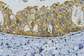
HER2 (ERBB2), CD117 (KIT) and TROP2 (TACSTD2) are surface and receptor proteins with established roles as tumour biomarkers, each tied to a different tumour context. HER2 gene amplification occurs in 18 to 20 percent of breast cancers and remains one of the most established prognostic and predictive biomarkers in solid tumour research.2 CD117 drives proliferation and survival signalling through the AKT and MAPK pathways, and strong CD117 expression is a defining diagnostic feature of gastrointestinal stromal tumours (GISTs). TROP2 is a calcium signal transducer overexpressed on the surface of many epithelial tumours and is the target of antibody-drug conjugates in development for breast and urothelial cancers.


Monoclonal Antibodies for Immunology Research
CD4, CD25 and CD69 are used together to characterise T cell identity and activation state by flow cytometry. CD4 acts as a coreceptor for MHC class II on helper T cells, recruiting the kinase LCK to the T cell receptor complex to initiate downstream signalling. CD25, the IL-2 receptor alpha chain, is expressed on activated T and B cells and is used alongside CD4 to identify regulatory T cell populations. CD69 is rapidly expressed on the surface of T cells, B cells and NK cells upon activation and is used as an early activation marker in immune monitoring assays.


Monoclonal Antibodies for Neuroscience Research
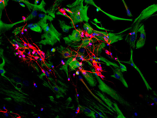
GFAP, NeuN and MAP2 mark distinct compartments of neural tissue and are used together to separate glial from neuronal populations in brain sections. GFAP is an intermediate filament protein expressed in astrocytes and is the standard marker for astrocytic processes. NeuN, encoded by RBFOX3, is localised largely to neuronal nuclei and is used to label post-mitotic neurons. MAP2 is a microtubule-associated protein restricted to neuronal dendrites and cell bodies, distinguishing neuronal processes from axons and glia.


Monoclonal Antibodies by Application Area
Beyond the featured research areas above, our mouse and rabbit monoclonal antibodies cover targets across signal transduction, extracellular matrix biology, hemostasis, vesicle-mediated transport and metabolic regulation. Each table below lists individual targets together with the host species available for that clone — expand a target to see specific clones.
Signal Transduction
Antibodies against receptor tyrosine kinases, MAPK pathway components and transcription factors, supporting studies of cell growth, differentiation and survival signalling.
| Target | Host | Application | Notes | Clones |
|---|---|---|---|---|
| FGFR | Mouse | Kinase activation & inhibitor screens | RTK family driving proliferation and survival | |
| EGFR | Mouse | Cell signalling & growth assays | Study ligand binding, phosphorylation and inhibition in cancer models | |
| EGFR | Rabbit | Cell signalling & inhibitor screening | Key oncology target driving proliferation | |
| HER2 | Mouse | Tumour profiling & antibody binding studies | Monitor overexpression in aggressive cancers | |
| NRP2 | Mouse | Angiogenesis & migration assays | VEGF co-receptor mediating vascular and tumour invasion pathways | |
| SEMA4D | Rabbit | Cell migration & immune modulation | Membrane semaphorin regulating immune activation pathways | |
| c-Kit/CD117 | Rabbit | Stem cell & oncology signalling | RTK with a role in hematopoiesis and cell survival | |
| Notch1 | Rabbit | Developmental & differentiation assays | Transmembrane receptor directing T cell fate and cell-cell communication | |
| ERK1/2 | Rabbit | MAPK pathway | Effector kinase in the RAS-RAF-MEK-ERK cascade | |
| MEK1/2 | Rabbit | Pathway activation & inhibitor profiling | MAPK pathway analysis | |
| FOXO3 | Rabbit | Transcription & apoptosis assays | Regulates metabolism, survival and stress response | |
| SRC | Rabbit | Kinase activity & adhesion assays | Cytoskeletal dynamics and proliferation | |
| Phospho-SRC | Rabbit | Activation & signalling flux assays | Marker for active signalling and kinase regulation | |
| Phospho-NFκB (p65) | Rabbit | Inflammatory signalling & transcription | Cytokine response and pathway flux assessment | |
| CAMK2α | Rabbit | Neuronal signalling & plasticity studies | Key player in synaptic plasticity and memory formation | |
Extracellular Matrix & Cell Adhesion
Antibodies against structural ECM glycoproteins, cytoskeletal proteins and adhesion complexes, supporting studies of tissue integrity, matrix remodelling and epithelial-mesenchymal transition.
| Target | Host | Application | Notes | Clones |
|---|---|---|---|---|
| Laminin | Mouse | ECM modelling & cell adhesion assays | Key basement membrane glycoprotein; supports adhesion, differentiation and migration | |
| E-cadherin | Mouse | EMT, cell polarity & cell-cell junctions | Key epithelial adhesion molecule; monitor loss during EMT or cancer invasion models | |
| ICAM-1 | Mouse | Immune cell trafficking & inflammation models | Endothelial adhesion receptor; study leukocyte binding and transendothelial migration | |
| Vinculin | Rabbit | Focal adhesion | Crucial for adhesion and force transmission studies | |
| Filamin A | Rabbit | Cytoskeleton & migration studies | Actin-crosslinking protein regulating cell shape, motility and signalling | |
| Alpha Actin | Rabbit | Muscle contraction & cytoskeletal assays | Core structural microfilament protein; essential for contractility and cellular dynamics | |
| Pan Cadherin | Rabbit | Cell-cell adhesion & EMT models | Broad reactivity to E-, N- and P-Cadherins; tracks epithelial integrity and transition | |
| MMP12 | Rabbit | ECM remodelling & inflammation studies | Important in tissue injury and repair pathway research | |
Hemostasis
Antibodies supporting coagulation and platelet biology research.
| Target | Host | Application | Notes | Clones |
|---|---|---|---|---|
| VWF | Mouse | Hemostasis & thrombosis models | Key glycoprotein contributing to platelet function | |
Vesicle-Mediated Transport
Antibodies for tracing intracellular trafficking, endocytosis and vesicle fusion across the stages of cargo movement between organelles.
| Target | Host | Application | Notes | Clones |
|---|---|---|---|---|
| LAMP1 | Mouse | Lysosome & autophagy studies | Monitor lysosomal activity and autophagy in cells | |
| Syntaxin 6 | Rabbit | Endosome trafficking & fusion assays | T-SNARE protein critical for endosomal/Golgi transport and vesicle fusion events | |
| Phospho Caveolin 1 | Mouse | Membrane dynamics & signalling | Involved in regulation of endocytosis, lipid signalling and receptor-mediated pathways | |
| DYNLL1 | Rabbit | Microtubule transport | Crucial for retrograde cargo trafficking and cytoskeletal transport dynamics | |
| STUB1 | Rabbit | Protein degradation & chaperone quality control | E3 ubiquitin ligase linking HSP70/HSP90 to proteasomal turnover | |
| EXOC2 | Rabbit | Exocytosis assays | Mediates vesicle docking and polarised secretion at the plasma membrane | |
| Transferrin | Rabbit | Endocytosis & receptor trafficking assays | Marker for receptor-mediated uptake | |
Metabolism & Cellular Stress Response
Antibodies against metabolic and regulatory enzymes, supporting studies of lipid metabolism, energy balance and cardiovascular disease.
| Target | Host | Application | Notes | Clones |
|---|---|---|---|---|
| PCSK9 | Mouse | Cholesterol regulation & cardiovascular models | Secreted enzyme mediating LDL receptor degradation; regulates plasma cholesterol and lipid metabolism | |
| MMP9 | Mouse | Atherosclerosis studies | Linked to vascular remodelling and cholesterol homeostasis; used in inflammation and ECM assays | |
Available Conjugation Types
Direct Detection & Multiplexing with Conjugated Antibodies
Conjugated primary antibodies carry a fluorophore or enzyme label directly on the antibody, enabling one-step direct detection of an antigen without a secondary antibody incubation and its associated wash steps.3 Because the label binds the extracellular epitope directly, these formats are particularly important for flow cytometry, where direct labelling is generally preferred for multi-parameter panels: it requires fewer processing steps and reduces the risk of cross-reactivity between antibodies raised in the same host species.3
- HRP: colourimetric or chemiluminescent signal for ELISA and western blotting.
- Biotin: signal amplification for low-expression targets.
- Fluorescent dyes: Alexa Fluor™, AbFluor™, FITC, PE and APC for multicolour flow cytometry and immunofluorescence.
- Tandem dyes (PE/Cyanine 7, PerCP/Cyanine 5.5): wide spectral separation from a single excitation laser for deep multi-target panels.
Flexible Assay Design with Unconjugated Antibodies
Unconjugated formats suit classic two-step indirect detection and provide high specificity when paired with a validated secondary antibody.
- Workflow flexibility: use one primary antibody across multiple applications by changing the labelled secondary.
- Enhanced sensitivity: indirect detection with a labelled secondary provides an amplified signal for low-expression targets.
- Bulk quantities available: suited to in-house conjugation or large-scale screening at high-concentration, unconjugated format.
Click a tab above to compare conjugated and unconjugated formats.
Learning and Support
Choosing between monoclonal and polyclonal antibodies?
Read our guide comparing the two formats: Monoclonal versus polyclonal antibody — which to choose?
Not sure which clone or host species fits your assay? Talk to us and our team can help you select the right product.
Looking for a Specific Target?
Browse the full range of 39,843 monoclonal antibodies, or contact us if a specific target is not listed.
Browse Monoclonal Antibodies1. Scholzen T, Gerdes J. The Ki-67 protein: from the known and the unknown. J Cell Physiol. 2000;182(3):311-322.
2. Bregni G, Beelen P, Kirilovsky A, et al. Targeting ERBB2 (HER2) Amplification Identified by Next-Generation Sequencing in Patients With Advanced or Metastatic Solid Tumors. JCO Precis Oncol. 2019;3:1-9.
3. Perfetto SP, Chattopadhyay PK, Roederer M. Seventeen-colour flow cytometry: unravelling the immune system. Nat Rev Immunol. 2004;4(8):648-655.
4. Bradbury A, Plückthun A. Reproducibility: Standardize antibodies used in research. Nature. 2015;518(7537):27-29.
5. Frenzel A, Hust M, Schirrmann T. Expression of recombinant antibodies. Front Immunol. 2013;4:217.