
Loading Control Antibodies
Our loading control range targets the housekeeping proteins used to normalise western blot signal across cytosolic, nuclear, membrane and mitochondrial fractions. GAPDH, Beta-Actin and Tubulin cover whole-cell and cytoplasmic lysates, Lamin A/C and PCNA cover nuclear fractions, and Na/K-ATPase and COX IV cover membrane and mitochondrial preparations. Monoclonal and polyclonal formats are available, with unconjugated, HRP and DyLight fluorescent conjugate options for a number of targets.
Browse Loading Control AntibodiesWhat Are Loading Control Antibodies?
Loading control antibodies target proteins with consistently high, ubiquitous expression across most cell types, tissues and species. Detecting one of these housekeeping proteins on the same blot as a target protein confirms that differences in band intensity between lanes reflect genuine biological change rather than inconsistencies in sample preparation, protein quantification or gel transfer.
This matters most when comparing samples across different treatment conditions, time points or cell lines, where small variations in loading can otherwise be read as meaningful biological differences.
The most widely used loading controls target cytosolic proteins such as GAPDH, Beta-Actin and Tubulin. Histone H3 and Lamin A/C are commonly used for nuclear fractions, and Na/K-ATPase and COX IV serve as controls for membrane and mitochondrial fractions.
Choosing the Right Loading Control
Three criteria guide the choice of control. Click a card for more on each one.
Common Pitfalls to Avoid
GAPDH and Alpha-Tubulin: The Two Most Searched Loading Controls
GAPDH and Alpha-Tubulin account for the largest share of search interest across our loading control range. GAPDH is a glycolytic enzyme present at high, stable levels in most cell types outside hypoxic or metabolically altered conditions.1 Alpha-Tubulin is a core structural component of the microtubule cytoskeleton and runs at 50 kDa, a molecular weight well separated from both GAPDH and Beta-Actin, making it a useful alternative when a target protein overlaps with either.

Anti-GAPDH Antibody [2B8] (STJ96931)
A mouse monoclonal antibody raised against amino acids 200 to 300 of human GAPDH, reactive across human, rat, mouse and monkey samples plus 9 further species. Validated for western blot, immunofluorescence and immunohistochemistry, detecting the protein at its expected 37 kDa.
View product →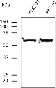
Anti-TUBA4A / Alpha-Tubulin Antibody (400aa C-Term) (STJ140051)
A goat polyclonal antibody raised against the 400 amino acid C-terminal region of Alpha-Tubulin, reactive across 17 species. Validated for western blot and immunofluorescence, detecting the protein at its expected 50 kDa.
View product →GAPDH Loading Controls
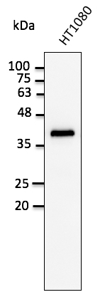
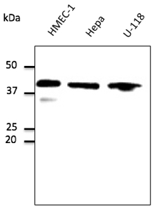
GAPDH (glyceraldehyde-3-phosphate dehydrogenase) is a glycolytic enzyme expressed at high levels across most mammalian cell types, and it runs at 37 kDa, well separated from most other cytosolic and membrane targets. It detects reliably by western blot, immunofluorescence and immunohistochemistry. Because glycolytic flux changes with oxygen availability and metabolic state, GAPDH expression varies between tissue types and is affected by hypoxia, so an alternative control is more appropriate for these conditions.1
GAPDH Also Available with DyLight Conjugates
For secondary-free fluorescence western blotting, GAPDH is also available pre-conjugated to DyLight 405, 488, 550 and 633. This removes the secondary antibody incubation step from a multiplex blotting workflow and reduces non-specific background.
Beta-Actin (ACTB) Loading Controls
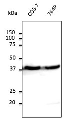
Beta-Actin is a cytoskeletal microfilament protein expressed across most cell types and detected at 42 kDa, separated from GAPDH by 5 kDa. It remains one of the most widely cited loading controls in the published literature. Actin expression can differ between tissue types and change during cytoskeletal reorganisation or cell differentiation, so it is not the most reliable single control for studies where either process is the variable under investigation.2


Tubulin Loading Controls
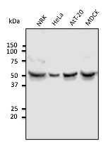
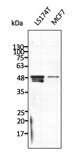
Alpha- and Beta-Tubulin are structural components of the microtubule cytoskeleton, both detected at 50 kDa, a molecular weight that separates them from GAPDH and Beta-Actin on the same blot. Tubulin expression can be affected by antimitotic or microtubule-targeting drug treatment, which is worth accounting for in drug-response studies.2

Nuclear Loading Controls
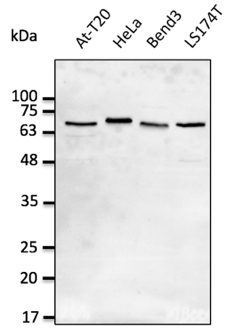
Cytosolic controls such as GAPDH and Beta-Actin are not appropriate for nuclear extracts or subcellular fractionation experiments. Lamin A/C, a nuclear envelope intermediate filament protein, runs at 74 kDa, separated from most cytosolic housekeeping proteins. PCNA (proliferating cell nuclear antigen), at 29 kDa, marks nuclear content in actively dividing cells, but should be avoided in experiments involving DNA damage or cell cycle arrest, where PCNA levels themselves change.

Histone H3 in the Wider Nuclear Range
Histone H3 is a further nuclear loading control target in our wider catalogue, alongside Lamin A/C and PCNA above. As a core histone bound to chromatin, Histone H3 levels change across the cell cycle as DNA replicates, so it should be avoided as a sole control in cell cycle studies.
Membrane and Mitochondrial Loading Controls
Membrane and mitochondrial fractions need controls distinct from the cytosolic and nuclear targets above. Na/K-ATPase is a plasma membrane ion pump that maintains the sodium and potassium gradient across the cell membrane, catalysing ATP hydrolysis to drive the exchange. COX IV is a subunit of Complex IV in the mitochondrial electron transport chain, the terminal enzyme that reduces oxygen to water during oxidative phosphorylation. Both are established controls for these compartments, and both fraction categories draw consistent search interest for our range.


Cytoskeletal and Adhesion Loading Controls
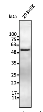
Vimentin, an intermediate filament protein, and Vinculin, a focal adhesion protein that links the actin cytoskeleton to the plasma membrane, are useful controls in mesenchymal, stromal and fibroblast cell models where the standard cytosolic controls are not always the best fit. Vimentin runs at 54 kDa and Vinculin at 117 kDa, both well separated from GAPDH and Beta-Actin on the same blot.

Loading Control Selection Guide
A loading control's molecular weight should sit at least 5 kDa from the protein of interest, and its expression should be stable under the experimental conditions in use. This table summarises the targets covered across the sections above.
| Target | Fraction | MW | Notes | Browse |
|---|---|---|---|---|
| GAPDH | Cytosolic | 37 kDa | Most widely used; avoid in hypoxia or altered metabolic state studies | Browse |
| Beta-Actin (ACTB) | Cytosolic | 42 kDa | Widely cited; not suited to cell growth or cytoskeletal reorganisation studies | Browse |
| Alpha/Beta-Tubulin | Cytosolic | 50 kDa | Good option when target is not near 50 kDa; may vary with antimitotic drug treatment | Browse |
| Vimentin (VIM) | Cytoskeletal | 54 kDa | Intermediate filament marker; useful in mesenchymal and fibroblast cell studies | Browse |
| Vinculin (VCL) | Adhesion | 117 kDa | Focal adhesion marker; distinct molecular weight from all cytosolic controls above | Browse |
| Lamin A/C (LMNA) | Nuclear | 74 kDa | Nuclear envelope marker; useful for nuclear structure or laminopathy studies | Browse |
| PCNA | Nuclear | 29 kDa | DNA replication marker; not suitable for non-proliferating cells or DNA damage experiments | Browse |
| Na/K-ATPase (ATP1A1) | Membrane | 113 kDa | Plasma membrane control; useful for membrane fraction validation | Browse |
| COX IV (COX4I1) | Mitochondrial | 17 kDa | Inner mitochondrial membrane control for mitochondrial fraction experiments | Browse |
Why Choose Loading Control Antibodies from Our Range
Monoclonal and Polyclonal Options
Both formats are available for key targets. Monoclonals offer high specificity and lot-to-lot consistency, and polyclonals give broad multi-epitope coverage for stronger signal detection.
Broad Species Reactivity
Most antibodies in the STJ140 goat polyclonal range are reactive across up to 17 species, including human, mouse, rat, bovine, porcine, rabbit, sheep and simian.
Multiple Subcellular Compartments
Coverage spans cytosolic, nuclear, membrane and mitochondrial fractions, so the right control is available regardless of which compartment the target protein occupies.
Full Datasheets
Each antibody is supplied with a datasheet. Where available, representative western blot images, dilution recommendations and related reagents are included.
Unconjugated, HRP and DyLight Conjugate Formats
Unconjugated Primary Antibodies
Used with a compatible secondary antibody for detection. This gives flexibility across secondary formats and detection systems including ECL, fluorescence and chromogenic methods, and suits assays where a labelled secondary is already in use for the target protein.
HRP-Conjugated Loading Controls
HRP-conjugated loading control antibodies allow direct detection without a secondary antibody step, reducing background and simplifying the western blot workflow for high-throughput or repeat use.
DyLight Fluorescent Conjugates
Pre-conjugated to DyLight 405, 488, 550 and 633. Suited to multiplexed fluorescence western blotting alongside a target antibody in a different channel, without secondary antibody cross-reactivity between the target and control.
Click a tab above to compare conjugate formats.
Western Blot and Loading Control Resources
Loading Control Antibody Guide
Read the Guide →Background on loading control selection, subcellular localisation and common pitfalls in western blot normalisation.
Western Blot Protocol
View Protocol →Step-by-step protocol covering sample preparation, SDS-PAGE, transfer, blocking and detection.
Western Blot Troubleshooting
Troubleshooting Guide →Diagnose and fix common issues including high background, weak signal, incorrect band size and uneven loading.
Looking for a Specific Loading Control?
Browse the full loading control range, including unconjugated, HRP and DyLight-conjugated antibodies for GAPDH, Beta-Actin, Tubulin, Histone H3, PCNA, Lamin A/C, Na/K-ATPase, COX IV, Vimentin and Vinculin.
Browse Loading Control Antibodies1. Barber RD, Harmer DW, Coleman RA, Clark BJ. GAPDH as a housekeeping gene: analysis of GAPDH mRNA expression in a panel of 72 human tissues. Physiol Genomics. 2005;21(3):389-395.
2. Eaton SL, Roche SL, Llavero Hurtado M, et al. Total protein analysis as a reliable loading control for quantitative fluorescent western blotting. PLoS ONE. 2013;8(8):e72457.