• Western blot analysis of extracts from HeLa cells, using Na+/K+-ATPase rabbit monoclonal antibody (STJ11101388) at 1:50000 dilution. Membrane protein extract isolated from Hela cells. Secondary antibody: HRP Goat Anti-rabbit IgG (H+L) (STJS000856) at 1:10000 dilution. Lysates/proteins: 25 Mu g per lane. Blocking buffer: 3% non-fat dry milk in TBST. Detection: ECL Basic Kit. Exposure time: 30s.
  • Western blot analysis of various lysates, using Na+/K+-ATPase rabbit monoclonal antibody (STJ11101388) at 1:50000 dilution. Membrane protein extract isolated from Hela cells. Secondary antibody: HRP Goat Anti-rabbit IgG (H+L) (STJS000856) at 1:10000 dilution. Lysates/proteins: 25 Mu g per lane. Blocking buffer: 3% non-fat dry milk in TBST. Detection: ECL Basic Kit. Exposure time: 30s.
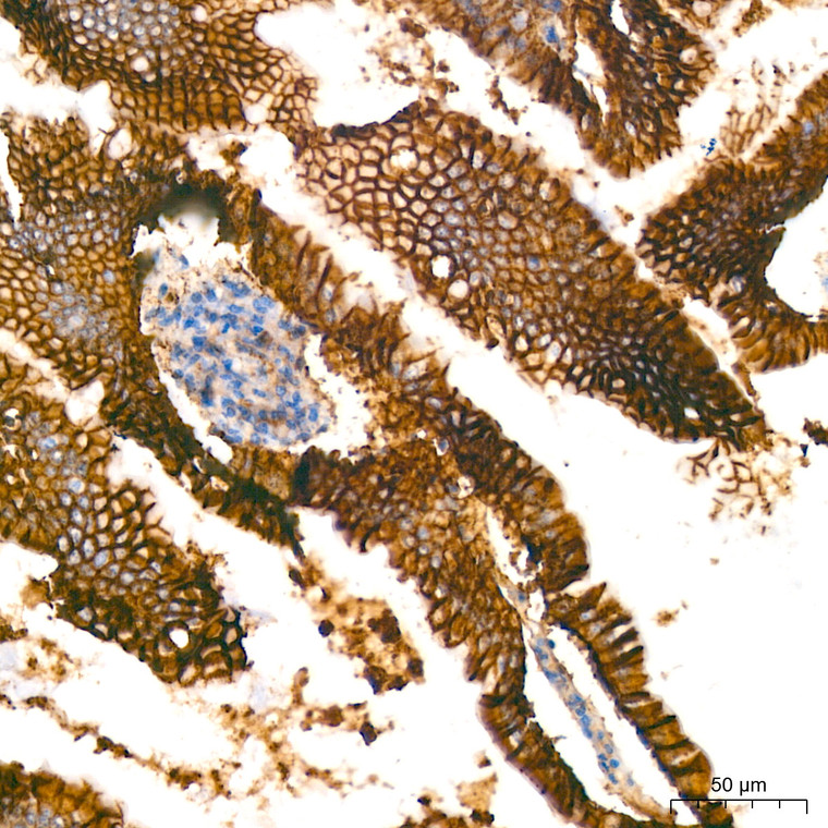
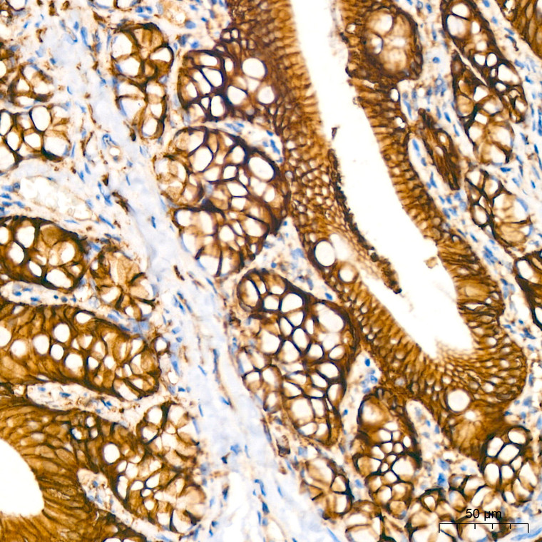
  • Immunohistochemistry analysis of paraffin-embedded human colon using Na+/K+-ATPase rabbit monoclonal antibody (STJ11101388) at dilution of 1:400 (40x lens). Perform high pressure antigen retrieval with 10 mM citrate buffer pH 6. 0 before commencing with immunohistochemistry staining protocol.
  • Immunohistochemistry analysis of paraffin-embedded human kidney using Na+/K+-ATPase rabbit monoclonal antibody (STJ11101388) at dilution of 1:400 (40x lens). Perform high pressure antigen retrieval with 10 mM citrate buffer pH 6. 0 before commencing with immunohistochemistry staining protocol.
  • Immunohistochemistry analysis of paraffin-embedded human thyroid cancer using Na+/K+-ATPase rabbit monoclonal antibody (STJ11101388) at dilution of 1:400 (40x lens). Perform high pressure antigen retrieval with 10 mM citrate buffer pH 6. 0 before commencing with immunohistochemistry staining protocol.
  • Immunohistochemistry analysis of paraffin-embedded human liver using Na+/K+-ATPase rabbit monoclonal antibody (STJ11101388) at dilution of 1:200 (40x lens). Perform high pressure antigen retrieval with 10 mM citrate buffer pH 6. 0 before commencing with immunohistochemistry staining protocol.
  • Immunohistochemistry analysis of paraffin-embedded mouse colon using Na+/K+-ATPase rabbit monoclonal antibody (STJ11101388) at dilution of 1:400 (40x lens). Perform high pressure antigen retrieval with 10 mM citrate buffer pH 6. 0 before commencing with immunohistochemistry staining protocol.
  • Immunohistochemistry analysis of paraffin-embedded rat colon using Na+/K+-ATPase rabbit monoclonal antibody (STJ11101388) at dilution of 1:400 (40x lens). Perform high pressure antigen retrieval with 10 mM citrate buffer pH 6. 0 before commencing with immunohistochemistry staining protocol.
  • Confocal imaging of human colon using Na+/K+-ATPase rabbit monoclonal antibody (STJ11101388, at dilution of 1:100) (Red). DAPI was used for nuclear staining (blue). Objective: 40x. Perform high pressure antigen retrieval with 10 mM citrate buffer pH 6. 0 before commencing with IF staining protocol.

Anti-ATP1A1 antibody (1-100) [S8MR] (STJ11101388)

SKU:
STJ11101388

Shipping:
Free Shipping
Current Stock:
Host: Rabbit
Applications: WB/IHC/IF
Reactivity: Human/Mouse/Rat
Note: STRICTLY FOR FURTHER SCIENTIFIC RESEARCH USE ONLY (RUO). MUST NOT TO BE USED IN DIAGNOSTIC OR THERAPEUTIC APPLICATIONS.
Short Description: Rabbit monoclonal antibody anti-Na+/K+-ATPase (1-100) is suitable for use in Western Blot, Immunohistochemistry and Immunofluorescence research applications.
Clonality: Monoclonal
Clone ID: S8MR
Conjugation: Unconjugated
Isotype: IgG
Formulation: PBS with 0.02% Sodium Azide, 0.05% BSA, 50% Glycerol, pH7.3.
Purification: Affinity purification
Dilution Range: WB 1:10000-1:120000
IHC-P 1:100-1:500
IF/ICC 1:50-1:200
Storage Instruction: Store at-20°C for up to 1 year from the date of receipt, and avoid repeat freeze-thaw cycles.
Gene Symbol: ATP1A1
Gene ID: 476
Uniprot ID: AT1A1_HUMAN
Immunogen Region: 1-100
Immunogen: A synthetic peptide corresponding to a sequence within amino acids 1-100 of human Na+/K+-ATPase (P05023).
Immunogen Sequence: MGKGVGRDKYEPAAVSEQGD KKGKKGKKDRDMDELKKEVS MDDHKLSLDELHRKYGTDLS RGLTSARAAEILARDGPNAL TPPPTTPEWIKFCRQLFGGF
Post Translational Modifications Phosphorylation on Tyr-10 modulates pumping activity. Phosphorylation of Ser-943 by PKA modulates the response of ATP1A1 to PKC. Dephosphorylation by protein phosphatase 2A (PP2A) following increases in intracellular sodium, leading to increase catalytic activity.
Function This is the catalytic component of the active enzyme, which catalyzes the hydrolysis of ATP coupled with the exchange of sodium and potassium ions across the plasma membrane. This action creates the electrochemical gradient of sodium and potassium ions, providing the energy for active transport of various nutrients.
Protein Name Sodium/Potassium-Transporting Atpase Subunit Alpha-1
Na(+/K(+ Atpase Alpha-1 Subunit
Sodium Pump Subunit Alpha-1
Database Links Reactome: R-HSA-5578775
Reactome: R-HSA-936837
Reactome: R-HSA-9679191
Cellular Localisation Basolateral Cell Membrane
Multi-Pass Membrane Protein
Cell Membrane
Sarcolemma
Cell Projection
Axon
Melanosome
Identified By Mass Spectrometry In Melanosome Fractions From Stage I To Stage Iv
Alternative Antibody Names Anti-Sodium/Potassium-Transporting Atpase Subunit Alpha-1 antibody
Anti-Na(+/K(+ Atpase Alpha-1 Subunit antibody
Anti-Sodium Pump Subunit Alpha-1 antibody
Anti-ATP1A1 antibody

Information sourced from Uniprot.org

12 months for antibodies. 6 months for ELISA Kits. Please see website T&Cs for further guidance