
IHC Guaranteed Antibodies
Our IHC Guaranteed range comprises primary antibodies validated for immunohistochemistry in formalin-fixed paraffin-embedded (FFPE) tissue sections, each tested against positive control tissue with results published in the product datasheet. The range covers site-of-origin, lineage and proliferation markers across carcinoma, lymphoma, tumour suppressor, cytokeratin and melanoma research, all reactive to human tissue and backed by a one-year product guarantee.
Browse IHC Guaranteed AntibodiesTwo Clones Behind Routine Tumour Assessment
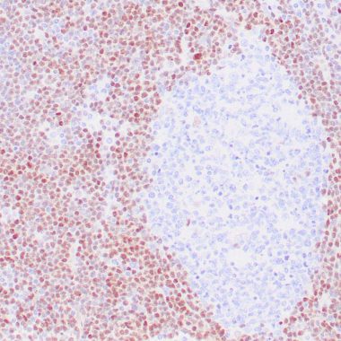
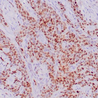
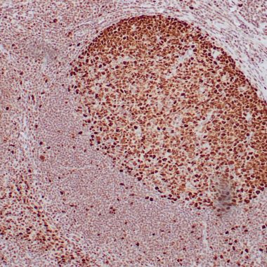
TP53 and MKI67 sit behind two of the most frequently reported IHC results in solid tumour pathology research: p53 protein accumulation studied as a surrogate marker for TP53 mutation status, and the Ki-67 proliferation index studied as a measure of tumour growth fraction. Both are available here as clones with an established track record in paraffin-embedded tissue, DO-7 for p53 and MIB-1 for Ki-67.
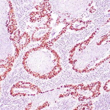
Anti-TP53 Antibody [DO-7] (STJ180207)
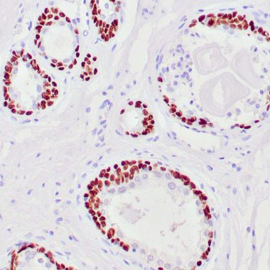
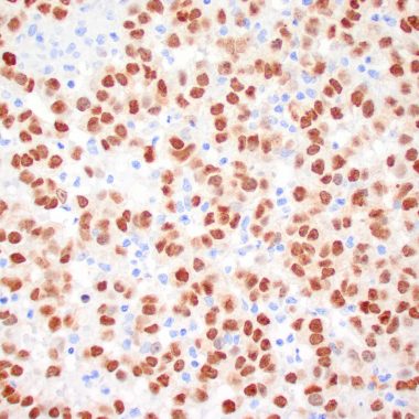
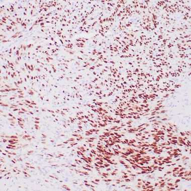
TP53 is among the most frequently altered genes in human cancer research models. Strong, diffuse nuclear p53 immunostaining is studied in ovarian carcinoma research as a surrogate marker for an underlying TP53 mutation, since this staining pattern is generally interpreted as indicating gene mutation.1 Clone DO-7 is validated for IHC on paraffin sections with heat-induced antigen retrieval.
View product →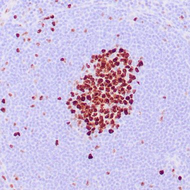
Anti-MKI67 Antibody [MIB-1] (STJ180156)
MIB-1 is considered the gold standard clone for Ki-67 evaluation, reported as the most frequently used antibody for this target with a long, validated track record in proliferation assessment.2 In breast cancer research, immunohistochemical assessment of Ki-67 positivity has become the most widely used method for comparing proliferation between tumour samples.3 This clone is validated for IHC in paraffin-embedded tissue.
View product →IHC Antibodies for Carcinoma & Adenocarcinoma
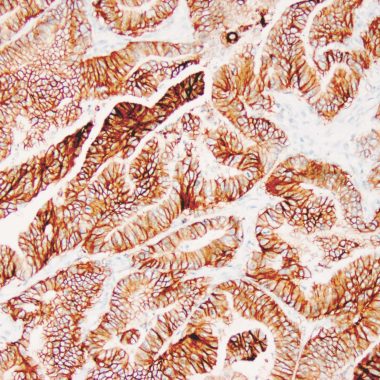
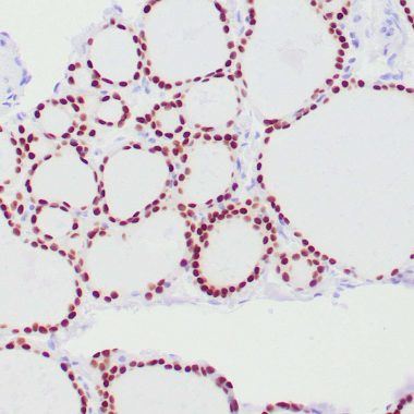
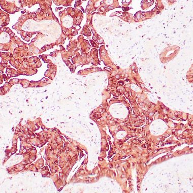
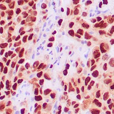
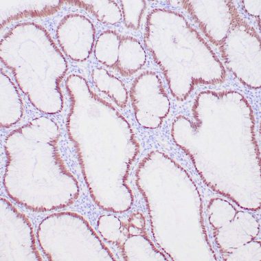
IHC antibodies for site-of-origin identification and lineage characterisation in paraffin-embedded carcinoma tissue. Commonly used to investigate tumour biology across lung, bladder, thyroid, kidney and gastrointestinal research models. Key targets include epithelial cell adhesion molecules, transcription factors and receptor tyrosine kinases whose expression patterns have been well characterised in primary carcinoma tissue. These IHC antibodies are widely used in studies examining tumour heterogeneity, metastatic spread and the molecular basis of carcinoma subtype differences.
IHC Antibodies for Lymphoma & Haematological Malignancy
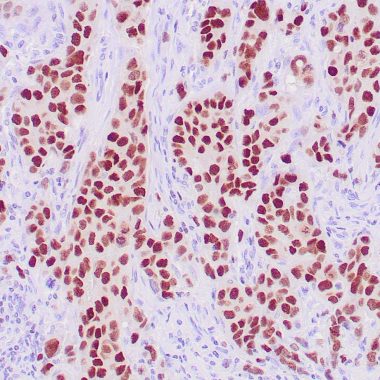
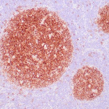
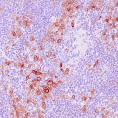
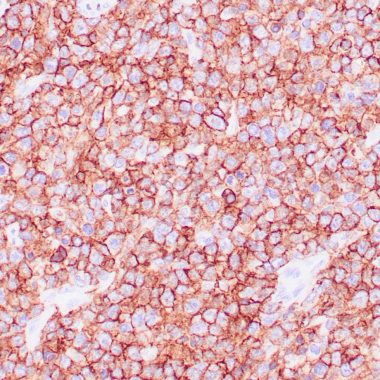
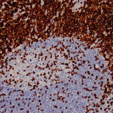
CD marker and lineage IHC antibodies for lymphoma subtype characterisation, leukaemia cell phenotyping and immune cell localisation in lymphoid tissue research. Validated in paraffin-embedded tonsil, lymph node and bone marrow sections. These IHC antibodies are widely used to investigate cell surface receptor expression, B and T cell identity, and the composition of the immune microenvironment in haematological malignancy models. They support research into lymphoma biology, clonal expansion and the functional properties of tumour-infiltrating immune populations.

IHC Antibodies for Tumour Suppressors & Cell Cycle
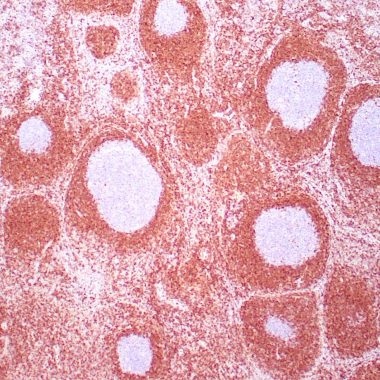
Validated IHC antibodies for studying cell cycle regulation, apoptosis and tumour suppressor protein expression in paraffin-embedded tissue sections. Used across cancer biology research to characterise proliferative activity and oncogene expression. These antibodies enable researchers to assess cell cycle progression markers, pro- and anti-apoptotic proteins including BCL2, and established tumour suppressors such as p16 and p63 in intact tissue architecture. They are commonly applied in studies examining growth control, DNA damage response and the molecular consequences of oncogene activation.
Cytokeratin IHC Antibodies & Epithelial Markers
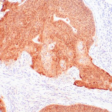
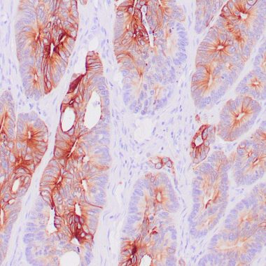
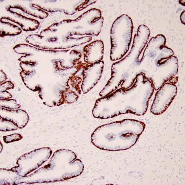
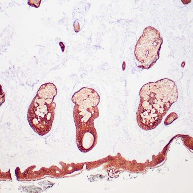
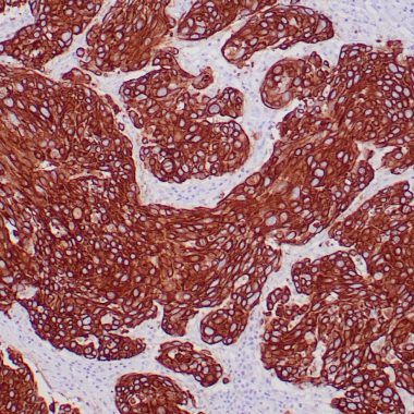
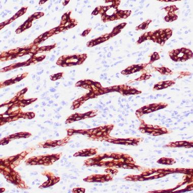
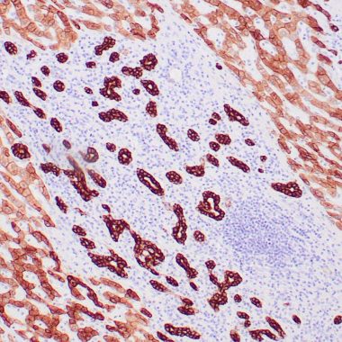
Cytokeratin IHC antibodies for characterising epithelial cell populations and identifying carcinoma origin in paraffin-embedded tissue. Includes pan-cytokeratin, high molecular weight cytokeratin and individual CK isoforms widely used in tissue-based research. Cytokeratin expression patterns are well-established tools for assessing epithelial differentiation state, studying epithelial-mesenchymal transition and distinguishing epithelial from stromal or haematological cell populations in mixed tissue sections. These IHC antibodies are routinely applied in studies of tumour invasion, metastasis and tissue organisation.
IHC Antibodies for Melanoma & Soft Tissue Tumours
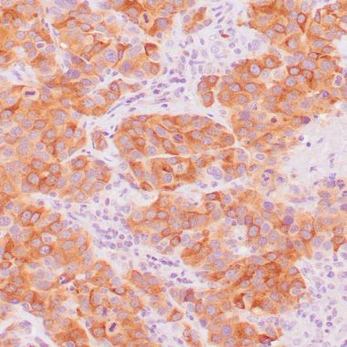
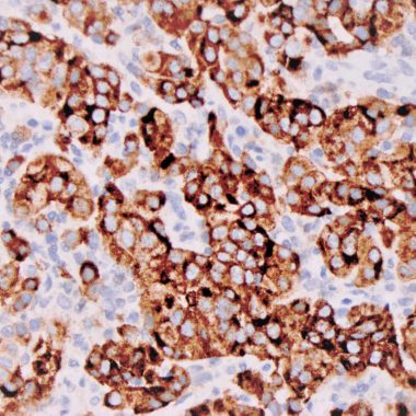
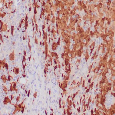
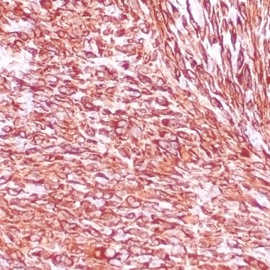
IHC antibodies for investigating melanocytic differentiation and soft tissue tumour biology in paraffin-embedded sections. Used in melanoma research to characterise marker expression and study tumour cell identity and differentiation state. Targets include established melanocytic markers such as Melan-A, HMB-45, MITF and S-100, as well as muscle lineage proteins including Desmin used to study myogenic tumour differentiation. These IHC antibodies are applied in research examining melanoma progression, neural crest lineage biology and the molecular basis of soft tissue tumour classification.
Looking for a Specific Target?
Browse our IHC Guaranteed antibodies, or contact us if a specific target is not listed.
Browse IHC Guaranteed Antibodies1. Yemelyanova A, Vang R, Kshirsagar M, Lu D, Marks MA, Shih IM, Kurman RJ. Immunohistochemical staining patterns of p53 can serve as a surrogate marker for TP53 mutations in ovarian carcinoma: an immunohistochemical and nucleotide sequencing analysis. Mod Pathol. 2011;24(9):1248-1253.
2. Zheng H, et al. Ki-67: a Promising Prognostic Marker in Early Breast Cancer. PMC9986372.
3. Dowsett M, Nielsen TO, A'Hern R, et al. Assessment of Ki67 in breast cancer: recommendations from the International Ki67 in Breast Cancer working group. J Natl Cancer Inst. 2011.