
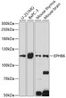


Anti-EPHB6 antibody (615-729) (STJ116928)
SPECIFICATIONS
ClonalityPolyclonal
HostRabbit
ConjugationUnconjugated
IsotypeIgG
General Information
| Short Description | Rabbit polyclonal EPHB6 (615-729) antibody for use in WB and ELISA in human and mouse samples. Datasheet included with dilution recommendations, and related reagents. |
| Applications | WB/ELISA |
| Host | Rabbit |
| Reactivity | Human/Mouse |
| Note | STRICTLY FOR FURTHER SCIENTIFIC RESEARCH USE ONLY (RUO). MUST NOT TO BE USED IN DIAGNOSTIC OR THERAPEUTIC APPLICATIONS. |
Product Properties
| Clonality | Polyclonal |
| Isotype | IgG |
| Conjugation | Unconjugated |
| Concentration | Lot specific |
| Purification | Affinity purification |
| Dilution Range | WB:1:500-1:2000ELISA:Recommended starting concentration is 1 Mu g/mL. Please optimize the concentration based on your specific assay requirements. |
| Formulation | PBS with 0.01% Thimerosal, 50% Glycerol, pH 7.3. |
| Storage Instruction | Store at-20°C for up to 1 year from the date of receipt, and avoid repeat freeze-thaw cycles. |
Target Information
| Gene Symbol | EPHB6 |
| Gene ID | 2051 |
| Uniprot ID | EPHB6_HUMAN |
| Immunogen Region | 615-729 |
| Immunogen Sequence | HFDQLVAAFDKMIRKPDTLQ AGGDPGERPSQALLTPVALD FPCLDSPQAWLSAIGLECYQ DNFSKFGLCTFSDVAQLSLE DLPALGITLAGHQKKLLHHI QLLQQHLRQQGSVEV |
| Specificity | Recombinant fusion protein containing a sequence corresponding to amino acids 615-729 of human EPHB6 (NP_001267724.2). |
Additional Info
| Tissue Specificity | Expressed in brain. Expressed in non invasive breast carcinoma cell lines (at protein level). Strong expression in brain and pancreas, and weak expression in other tissues, such as heart, placenta, lung, liver, skeletal muscle and kidney. Expressed in breast non invasive tumors but not in metastatic lesions. Isoform 3 is expressed in cell lines of glioblastomas, anaplastic astrocytomas, gliosarcomas and astrocytomas. Isoform 3 is not detected in normal tissues. |
| Post Translational Modifications | Ligand-binding increases phosphorylation on tyrosine residues. Phosphorylation on tyrosine residues is mediated by transphosphorylation by the catalytically active EPHB1 in a ligand-independent manner. Tyrosine phosphorylation of the receptor may act as a switch on the functional transition from cell adhesion/attraction to de-adhesion/repulsion. |
| Function | Kinase-defective receptor for members of the ephrin-B family. Binds to ephrin-B1 and ephrin-B2. Modulates cell adhesion and migration by exerting both positive and negative effects upon stimulation with ephrin-B2. Inhibits JNK activation, T-cell receptor-induced IL-2 secretion and CD25 expression upon stimulation with ephrin-B2. |
| Protein Name | Ephrin Type-B Receptor 6HepTyrosine-Protein Kinase-Defective Receptor Eph-6 |
| Database Links | Reactome: R-HSA-2682334Reactome: R-HSA-3928662Reactome: R-HSA-3928664Reactome: R-HSA-3928665 |
| Cellular Localisation | MembraneSingle-Pass Type I Membrane ProteinIsoform 3: Secreted |
| Alternative Antibody Names | Anti-Ephrin Type-B Receptor 6 antibodyAnti-Hep antibodyAnti-Tyrosine-Protein Kinase-Defective Receptor Eph-6 antibodyAnti-EPHB6 antibody |
Information sourced from Uniprot.org