| Host: |
Goat |
| Applications: |
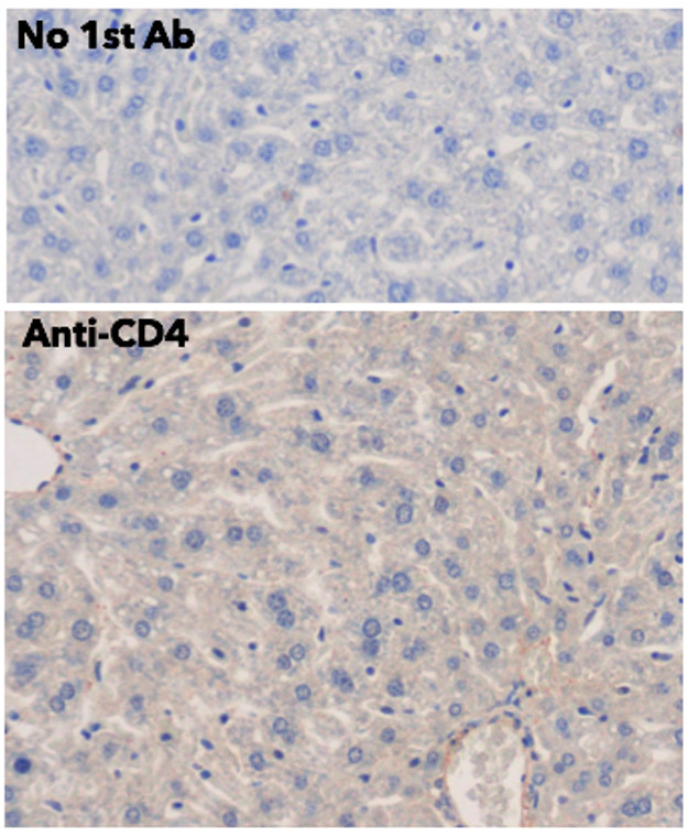
WB/IHC-F/IHC-P |
| Reactivity: |
Human/Rat/Mouse/Monkey/Canine |
| Note: |
STRICTLY FOR FURTHER SCIENTIFIC RESEARCH USE ONLY (RUO). MUST NOT TO BE USED IN DIAGNOSTIC OR THERAPEUTIC APPLICATIONS. |
| Short Description: |
Goat polyclonal antibody anti-CD4 molecule (50-235aa ECD) is suitable for use in Western Blot, Immunohistochemistry and Immunohistochemistry research applications. |
| Clonality: |
Polyclonal |
| Conjugation: |
Unconjugated |
| Isotype: |
IgG |
| Formulation: |
PBS, 20% Glycerol and 0.05% Sodium Azide. |
| Purification: |
This antibody is epitope-affinity purified from goat antiserum. |
| Concentration: |
3 mg/ml |
| Dilution Range: |
WB 1:500-1:2000IHC-F 1:250-1:1000IHC-P 1:250-1:1000Helsby MA Leader PM Fenn JR et al. BMC Cell Biol. 2014 Feb 14:15:6. PMID: 24528853 |
| Storage Instruction: |
For continuous use, store at 2-8 C for one-two days. For extended storage, store in-20 C freezer. Working dilution samples should be discarded if not used within 12 hours. |
| Gene Symbol: |
CD4 |
| Gene ID: |
920 |
| Uniprot ID: |
CD4_HUMAN |
| Immunogen Region: |
50-235aa ECD |
| Accession Number: |
ENSG00000010610 |
| Specificity: |
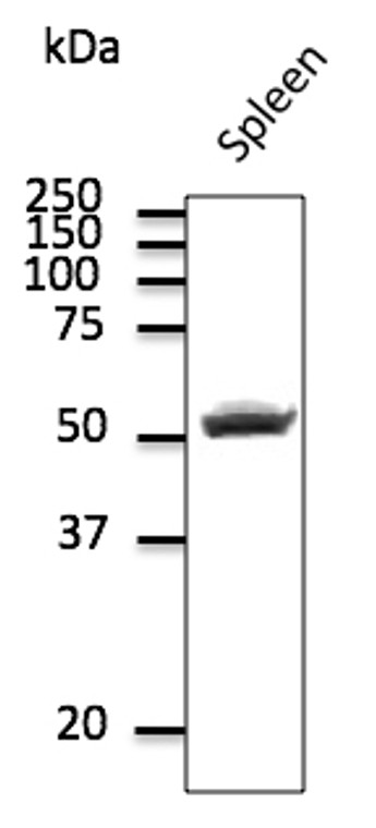
Using spleen lysate detects a 52 kDa band by Western blot. |
| Immunogen: |
Purified recombinant peptide derived from within 50 to 235 aa, corresponding to the external domain of human CD4 produced in E. coli. |
| Immunogen Sequence: |
FHWKNSNQIKILGNQGSFLT KGPSKLNDRADSRRSLWDQG NFPLIIKNLKIEDSDTYICE VSQLELQDSGTWTCTVLQNQ KKVEFKIDIVVLAFQKASSI VYKKEGEQVEFSFPLAFTVE VEDQKEEVQLLVFGLTANSD THLLQGQSLTLTLESPPGSS PSVQCRSPRGKNIQGGKTLS KLTGSG |
| Tissue Specificity | Highly expressed in T-helper cells. The presence of CD4 is a hallmark of T-helper cells which are specialized in the activation and growth of cytotoxic T-cells, regulation of B cells, or activation of phagocytes. CD4 is also present in other immune cells such as macrophages, dendritic cells or NK cells. |
| Post Translational Modifications | Palmitoylation and association with LCK contribute to the enrichment of CD4 in lipid rafts. Phosphorylated by PKC.phosphorylation at Ser-433 plays an important role for CD4 internalization. |
| Function | Integral membrane glycoprotein that plays an essential role in the immune response and serves multiple functions in responses against both external and internal offenses. In T-cells, functions primarily as a coreceptor for MHC class II molecule:peptide complex. The antigens presented by class II peptides are derived from extracellular proteins while class I peptides are derived from cytosolic proteins. Interacts simultaneously with the T-cell receptor (TCR) and the MHC class II presented by antigen presenting cells (APCs). In turn, recruits the Src kinase LCK to the vicinity of the TCR-CD3 complex. LCK then initiates different intracellular signaling pathways by phosphorylating various substrates ultimately leading to lymphokine production, motility, adhesion and activation of T-helper cells. In other cells such as macrophages or NK cells, plays a role in differentiation/activation, cytokine expression and cell migration in a TCR/LCK-independent pathway. Participates in the development of T-helper cells in the thymus and triggers the differentiation of monocytes into functional mature macrophages. (Microbial infection) Primary receptor for human immunodeficiency virus-1 (HIV-1). Down-regulated by HIV-1 Vpu. Acts as a receptor for Human Herpes virus 7/HHV-7. |
| Protein Name | T-Cell Surface Glycoprotein Cd4T-Cell Surface Antigen T4/Leu-3Cd Antigen Cd4 |
| Database Links | Reactome: R-HSA-1462054Reactome: R-HSA-167590Reactome: R-HSA-173107Reactome: R-HSA-180534Reactome: R-HSA-202424Reactome: R-HSA-202427Reactome: R-HSA-202430Reactome: R-HSA-202433Reactome: R-HSA-389948Reactome: R-HSA-449836Reactome: R-HSA-8856825Reactome: R-HSA-8856828 |
| Cellular Localisation | Cell MembraneSingle-Pass Type I Membrane ProteinLocalizes To Lipid RaftsRemoved From Plasma Membrane By Hiv-1 Nef Protein That Increases Clathrin-Dependent Endocytosis Of This Antigen To Target It To Lysosomal DegradationCell Surface Expression Is Also Down-Modulated By Hiv-1 Envelope Polyprotein Gp160 That Interacts WithAnd Sequesters Cd4 In The Endoplasmic Reticulum |
| Alternative Antibody Names | Anti-T-Cell Surface Glycoprotein Cd4 antibodyAnti-T-Cell Surface Antigen T4/Leu-3 antibodyAnti-Cd Antigen Cd4 antibodyAnti-CD4 antibody |
Information sourced from Uniprot.org
12 months for antibodies. 6 months for ELISA Kits. Please see website T&Cs for further guidance


