| Host: |
Rabbit |
| Applications: |
WB/IF |
| Reactivity: |
Human/Mouse/Rat |
| Note: |
STRICTLY FOR FURTHER SCIENTIFIC RESEARCH USE ONLY (RUO). MUST NOT TO BE USED IN DIAGNOSTIC OR THERAPEUTIC APPLICATIONS. |
| Short Description: |
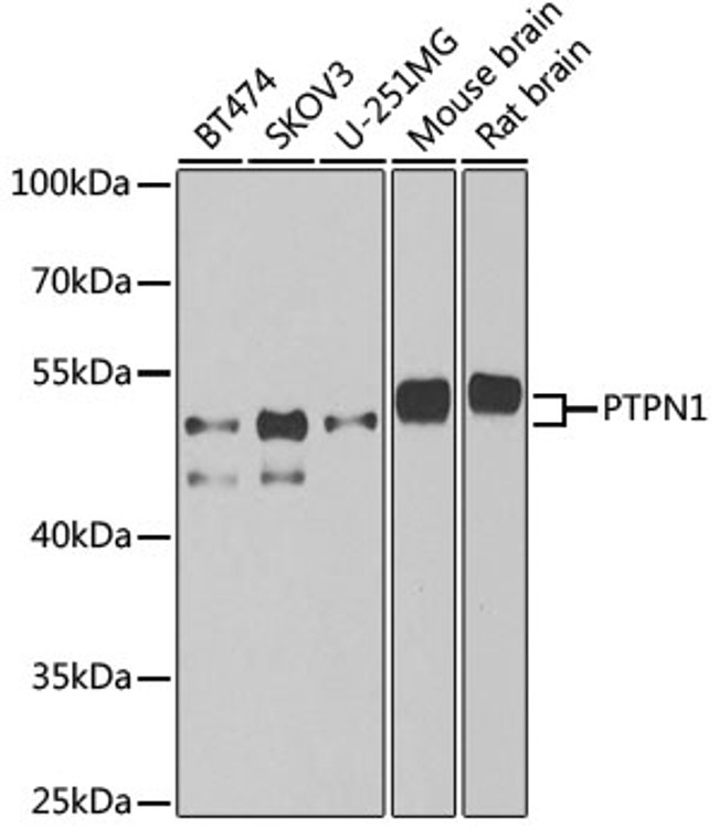
Rabbit polyclonal antibody anti-PTPN1 (281-435) is suitable for use in Western Blot and Immunofluorescence research applications. |
| Clonality: |
Polyclonal |
| Conjugation: |
Unconjugated |
| Isotype: |
IgG |
| Formulation: |
PBS with 0.05% Proclin300, 50% Glycerol, pH7.3. |
| Purification: |
Affinity purification |
| Dilution Range: |
WB 1:500-1:2000IF/ICC 1:50-1:200 |
| Storage Instruction: |
Store at-20°C for up to 1 year from the date of receipt, and avoid repeat freeze-thaw cycles. |
| Gene Symbol: |
PTPN1 |
| Gene ID: |
5770 |
| Uniprot ID: |
PTN1_HUMAN |
| Immunogen Region: |
281-435 |
| Immunogen: |
Recombinant fusion protein containing a sequence corresponding to amino acids 281-435 of human PTPN1 (NP_002818.1). |
| Immunogen Sequence: |
IMGDSSVQDQWKELSHEDLE PPPEHIPPPPRPPKRILEPH NGKCREFFPNHQWVKEETQE DKDCPIKEEKGSPLNAAPYG IESMSQDTEVRSRVVGGSLR GAQAASPAKGEPSLPEKDED HALSYWKPFLVNMCVATVLT AGAYLCYRFLFNSNT |
| Tissue Specificity | Expressed in keratinocytes (at protein level). |
| Post Translational Modifications | Oxidized on Cys-215.the Cys-SOH formed in response to redox signaling reacts with the alpha-amido of the following residue to form a sulfenamide cross-link, triggering a conformational change that inhibits substrate binding and activity. The active site can be restored by reduction. Ser-50 is the major site of phosphorylation as compared to Ser-242 and Ser-243. Activated by phosphorylation at Ser-50. S-nitrosylation of Cys-215 inactivates the enzyme activity. Sulfhydration at Cys-215 following endoplasmic reticulum stress inactivates the enzyme activity, promoting EIF2AK3/PERK activity. |
| Function | Tyrosine-protein phosphatase which acts as a regulator of endoplasmic reticulum unfolded protein response. Mediates dephosphorylation of EIF2AK3/PERK.inactivating the protein kinase activity of EIF2AK3/PERK. May play an important role in CKII- and p60c-src-induced signal transduction cascades. May regulate the EFNA5-EPHA3 signaling pathway which modulates cell reorganization and cell-cell repulsion. May also regulate the hepatocyte growth factor receptor signaling pathway through dephosphorylation of MET. |
| Protein Name | Tyrosine-Protein Phosphatase Non-Receptor Type 1Protein-Tyrosine Phosphatase 1bPtp-1b |
| Database Links | Reactome: R-HSA-354192Reactome: R-HSA-6807004Reactome: R-HSA-77387Reactome: R-HSA-877312Reactome: R-HSA-8849472Reactome: R-HSA-9022699Reactome: R-HSA-912694Reactome: R-HSA-982772 |
| Cellular Localisation | Endoplasmic Reticulum MembranePeripheral Membrane ProteinCytoplasmic SideInteracts With Epha3 At The Cell Membrane |
| Alternative Antibody Names | Anti-Tyrosine-Protein Phosphatase Non-Receptor Type 1 antibodyAnti-Protein-Tyrosine Phosphatase 1b antibodyAnti-Ptp-1b antibodyAnti-PTPN1 antibodyAnti-PTP1B antibody |
Information sourced from Uniprot.org
12 months for antibodies. 6 months for ELISA Kits. Please see website T&Cs for further guidance


