• Western blot analysis of extracts of K562 cells, using Phospho-STAT5-Y694 polyclonal antibody (STJ11101025) at 1:1000 dilution. K-562 cells were treated by ATP (5 mM) at 30 °C for 1 hour. Secondary antibody: HRP Goat Anti-rabbit IgG (H+L) at 1:10000 dilution. Lysates/proteins: 25ug per lane. Blocking buffer: 3% BSA. Detection: ECL Basic Kit. Exposure time: 1s.
  • Immunohistochemistry analysis of Phospho-STAT5-Y694 in paraffin-embedded mouse liver using Phospho-STAT5-Y694 Rabbit polyclonal antibody (STJ11101025) at dilution of 1:200 (40x lens). Perform microwave antigen retrieval with 10 mM Tris/EDTA buffer pH 9. 0 before commencing with immunohistochemistry staining protocol.
  • Western blot analysis of extracts of A-431 cells, using Phospho-STAT5-Y694 polyclonal antibody (STJ11101025) at 1:1000 dilution or STAT5B antibody (A12356). A-431 cells were treated by EGF (100 ng/mL) at 37 °C for 30 minutes after serum-starvation overnight. Secondary antibody: HRP Goat Anti-rabbit IgG (H+L) at 1:10000 dilution. Lysates/proteins: 25ug per lane. Blocking buffer: 3% BSA. Detection: ECL Basic Kit. Exposure time: 60s.
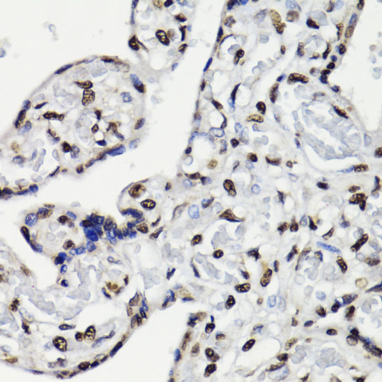
  • Immunohistochemistry analysis of Phospho-STAT5-Y694 in paraffin-embedded human placenta using Phospho-STAT5-Y694 Rabbit polyclonal antibody (STJ11101025) at dilution of 1:200 (40x lens). Perform microwave antigen retrieval with 10 mM Tris/EDTA buffer pH 9. 0 before commencing with immunohistochemistry staining protocol.
  • Immunohistochemistry of paraffin-embedded rat heart using Phospho-STAT5-Y694 antibody (STJ11101025) at dilution of 1:200 (40x lens).
  • Immunohistochemistry analysis of Phospho-STAT5-Y694 in paraffin-embedded rat heart using Phospho-STAT5-Y694 Rabbit polyclonal antibody (STJ11101025) at dilution of 1:200 (40x lens). Perform microwave antigen retrieval with 10 mM Tris/EDTA buffer pH 9. 0 before commencing with immunohistochemistry staining protocol.
  • Immunohistochemistry of paraffin-embedded human placenta using Phospho-STAT5-Y694 antibody (STJ11101025) at dilution of 1:200 (40x lens).
  • Western blot analysis of lysates from NIH/3T3 cells, using Phospho-STAT5-Y694 Rabbit polyclonal antibody (STJ11101025) at 1:400 dilution. NIH/3T3 cells were treated by Pervanadate (1 nM) at 37 °C for 30 minutes after serum-starvation overnight. Secondary antibody: HRP Goat Anti-Rabbit IgG (H+L) (STJS000856) at 1:10000 dilution. Lysates/proteins: 25 Mu g per lane. Blocking buffer: 3% nonfat dry milk in TBST. Detection: ECL Basic Kit. Exposure time: 1s.
  • Immunohistochemistry of paraffin-embedded mouse liver using Phospho-STAT5-Y694 antibody (STJ11101025) at dilution of 1:200 (40x lens).

Anti-Phospho-STAT5A/STAT5B-Y694 antibody (STJ11101025)

SKU:
STJ11101025

Shipping:
Free Shipping
Current Stock:
Host: Rabbit
Applications: WB/IHC
Reactivity: Human/Mouse/Rat
Note: STRICTLY FOR FURTHER SCIENTIFIC RESEARCH USE ONLY (RUO). MUST NOT TO BE USED IN DIAGNOSTIC OR THERAPEUTIC APPLICATIONS.
Short Description: Rabbit polyclonal antibody anti-Phospho-STAT5-Y694 is suitable for use in Western Blot and Immunohistochemistry research applications.
Clonality: Polyclonal
Conjugation: Unconjugated
Isotype: IgG
Formulation: PBS with 0.01% Thimerosal, 50% Glycerol, pH7.3.
Purification: Affinity purification
Dilution Range: WB 1:100-1:500
IHC-P 1:100-1:200
Storage Instruction: Store at-20°C for up to 1 year from the date of receipt, and avoid repeat freeze-thaw cycles.
Immunogen: A synthetic phosphorylated peptide around Y694 of human STAT5 (NP_003143.2/NP_036580.2).
Immunogen Sequence: DGYVK
Background The protein encoded by this gene belongs to a family of bifunctional proteins that are involved in both the synthesis and degradation of fructose-2, 6-bisphosphate, a regulatory molecule that controls glycolysis in eukaryotes. The encoded protein has a 6-phosphofructo-2-kinase activity that catalyzes the synthesis of fructose-2, 6-bisphosphate (F2, 6BP) , and a fructose-2, 6-biphosphatase activity that catalyzes the degradation of F2, 6BP. This protein is required for cell cycle progression and prevention of apoptosis. It functions as a regulator of cyclin-dependent kinase 1, linking glucose metabolism to cell proliferation and survival in tumor cells. Several alternatively spliced transcript variants encoding different isoforms have been found for this gene.

Information sourced from Uniprot.org

12 months for antibodies. 6 months for ELISA Kits. Please see website T&Cs for further guidance