• Confocal imaging of C2C12 cells using LXR Alpha Rabbit monoclonal antibody (STJ11101976, dilution 1:100) followed by a further incubation with Cy3 Goat Anti-Rabbit IgG (H+L) ( dilution 1:500) (Red). The cells were counterstained with Alpha-Tubulin Mouse monoclonal antibody ( dilution 1:400) followed by incubation with ABflo® 488-conjugated Goat Anti-Mouse IgG (H+L) antibody ( dilution 1:500) (Green). DAPI was used for nuclear staining (Blue). Objective: 100x.
  • Immunofluorescence analysis of NIH-3T3 cells using LXR Alpha Rabbit monoclonal antibody (STJ11101976) at dilution of 1:100 (40x lens). Secondary antibody: Cy3 Goat Anti-Rabbit IgG (H+L) at 1:500 dilution. Blue: DAPI for nuclear staining.
  • Immunofluorescence analysis of Hep G2 cells using LXR Alpha Rabbit monoclonal antibody (STJ11101976) at dilution of 1:100 (40x lens). Secondary antibody: Cy3 Goat Anti-Rabbit IgG (H+L) at 1:500 dilution. Blue: DAPI for nuclear staining.
  • Immunofluorescence analysis of C6 cells using LXR Alpha Rabbit monoclonal antibody (STJ11101976) at dilution of 1:100 (40x lens). Secondary antibody: Cy3 Goat Anti-Rabbit IgG (H+L) at 1:500 dilution. Blue: DAPI for nuclear staining.
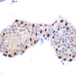
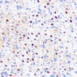
  • Immunohistochemistry analysis of LXR Alpha in paraffin-embedded mouse testis using LXR Alpha Rabbit monoclonal antibody (STJ11101976) at dilution of 1:100 (40x lens). Perform microwave antigen retrieval with 10 mM PBS buffer pH 7. 2 before commencing with immunohistochemistry staining protocol.
  • Immunohistochemistry analysis of LXR Alpha in paraffin-embedded human colon using LXR Alpha Rabbit monoclonal antibody (STJ11101976) at dilution of 1:100 (40x lens). Perform microwave antigen retrieval with 10 mM PBS buffer pH 7. 2 before commencing with immunohistochemistry staining protocol.
  • Immunohistochemistry analysis of LXR Alpha in paraffin-embedded rat ovary using LXR Alpha Rabbit monoclonal antibody (STJ11101976) at dilution of 1:100 (40x lens). Perform microwave antigen retrieval with 10 mM PBS buffer pH 7. 2 before commencing with immunohistochemistry staining protocol.
  • Western blot analysis of various lysates using LXR Alpha Rabbit monoclonal antibody (STJ11101976) at 1:1000 dilution. Secondary antibody: HRP Goat Anti-Rabbit IgG (H+L) (STJS000856) at 1:10000 dilution. Lysates/proteins: 25 Mu g per lane. Blocking buffer: 3% nonfat dry milk in TBST. Detection: ECL Basic Kit. Exposure time: 3s.

Anti-NR1H3 antibody (1-100) [S6MR] (STJ11101976)

SKU:
STJ11101976

Shipping:
Free Shipping
Current Stock:
Host: Rabbit
Applications: WB/IHC/IF
Reactivity: Human/Mouse/Rat
Note: STRICTLY FOR FURTHER SCIENTIFIC RESEARCH USE ONLY (RUO). MUST NOT TO BE USED IN DIAGNOSTIC OR THERAPEUTIC APPLICATIONS.
Short Description: Rabbit monoclonal antibody anti-LXR Alpha (1-100) is suitable for use in Western Blot, Immunohistochemistry and Immunofluorescence research applications.
Clonality: Monoclonal
Clone ID: S6MR
Conjugation: Unconjugated
Isotype: IgG
Formulation: PBS with 0.02% Sodium Azide, 0.05% BSA, 50% Glycerol, pH7.3.
Purification: Affinity purification
Dilution Range: WB 1:500-1:1000
IHC-P 1:50-1:200
IF/ICC 1:50-1:200
Storage Instruction: Store at-20°C for up to 1 year from the date of receipt, and avoid repeat freeze-thaw cycles.
Gene Symbol: NR1H3
Gene ID: 10062
Uniprot ID: NR1H3_HUMAN
Immunogen Region: 1-100
Immunogen: A synthetic peptide corresponding to a sequence within amino acids 1-100 of human LXR Alpha (Q13133).
Immunogen Sequence: MSLWLGAPVPDIPPDSAVEL WKPGAQDASSQAQGGSSCIL REEARMPHSAGGTAGVGLEA AEPTALLTRAEPPSEPTEIR PQKRKKGPAPKMLGNELCSV
Tissue Specificity Visceral organs specific expression. Strong expression was found in liver, kidney and intestine followed by spleen and to a lesser extent the adrenals.
Post Translational Modifications Ubiquitinated leading to its degradation by the proteasome.
Function Nuclear receptor that exhibits a ligand-dependent transcriptional activation activity. Interaction with retinoic acid receptor (RXR) shifts RXR from its role as a silent DNA-binding partner to an active ligand-binding subunit in mediating retinoid responses through target genes defined by LXRES. LXRES are DR4-type response elements characterized by direct repeats of two similar hexanuclotide half-sites spaced by four nucleotides. Plays an important role in the regulation of cholesterol homeostasis, regulating cholesterol uptake through MYLIP-dependent ubiquitination of LDLR, VLDLR and LRP8. Interplays functionally with RORA for the regulation of genes involved in liver metabolism. Induces LPCAT3-dependent phospholipid remodeling in endoplasmic reticulum (ER) membranes of hepatocytes, driving SREBF1 processing and lipogenesis. Via LPCAT3, triggers the incorporation of arachidonate into phosphatidylcholines of ER membranes, increasing membrane dynamics and enabling triacylglycerols transfer to nascent very low-density lipoprotein (VLDL) particles. Via LPCAT3 also counteracts lipid-induced ER stress response and inflammation, likely by modulating SRC kinase membrane compartmentalization and limiting the synthesis of lipid inflammatory mediators.
Protein Name Oxysterols Receptor Lxr-Alpha
Liver X Receptor Alpha
Nuclear Receptor Subfamily 1 Group H Member 3
Database Links Reactome: R-HSA-1989781
Reactome: R-HSA-383280
Reactome: R-HSA-4090294 Q13133-1
Reactome: R-HSA-8866427
Reactome: R-HSA-9029558
Reactome: R-HSA-9029569
Reactome: R-HSA-9031525
Reactome: R-HSA-9031528
Reactome: R-HSA-9623433
Reactome: R-HSA-9632974
Cellular Localisation Nucleus
Cytoplasm
Alternative Antibody Names Anti-Oxysterols Receptor Lxr-Alpha antibody
Anti-Liver X Receptor Alpha antibody
Anti-Nuclear Receptor Subfamily 1 Group H Member 3 antibody
Anti-NR1H3 antibody
Anti-LXRA antibody

Information sourced from Uniprot.org

12 months for antibodies. 6 months for ELISA Kits. Please see website T&Cs for further guidance