


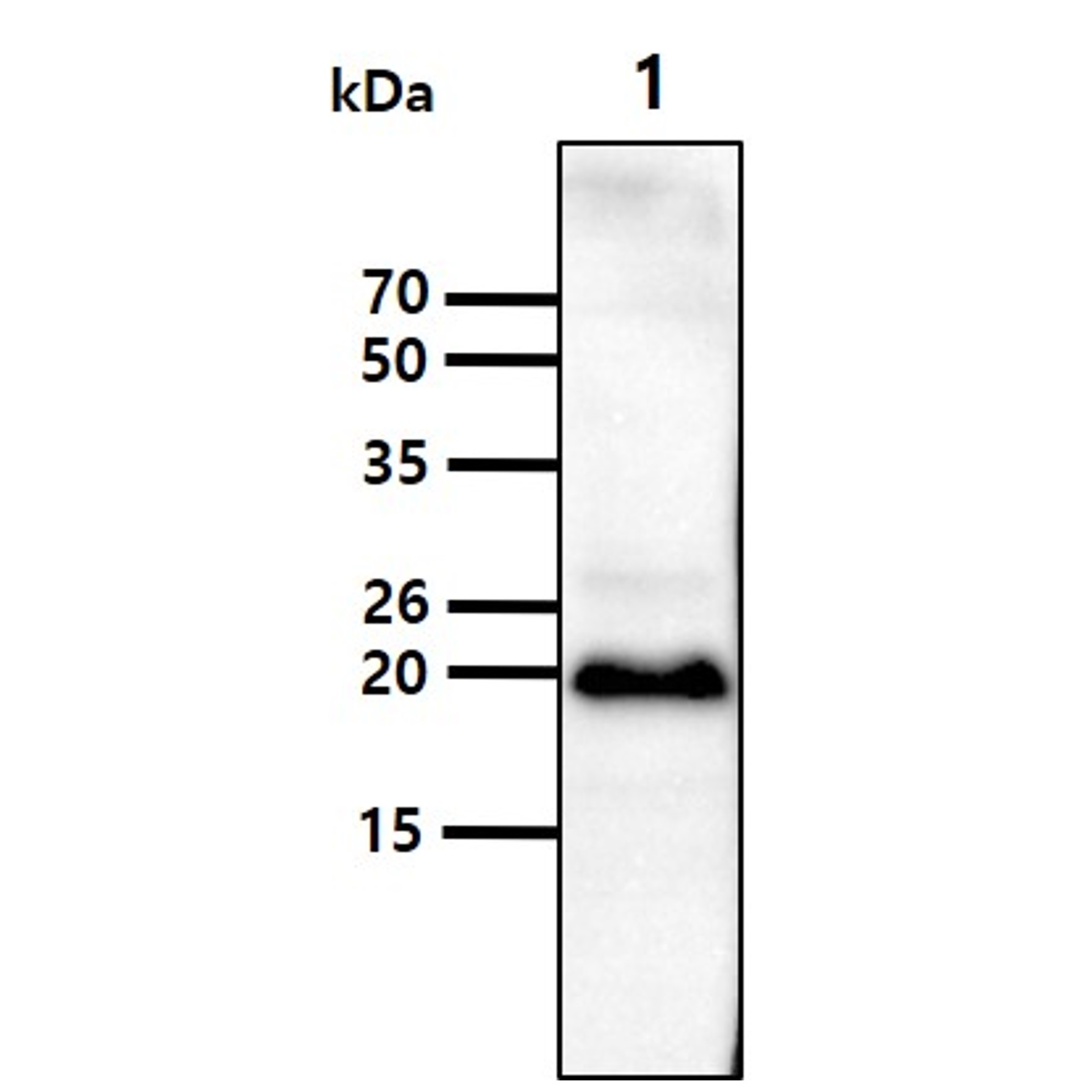
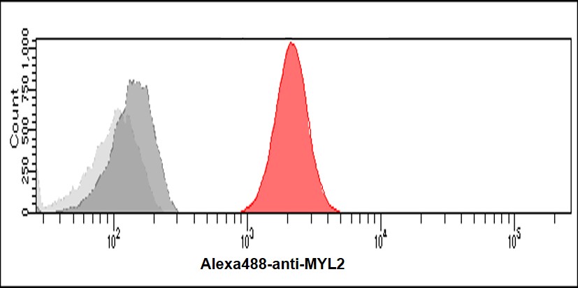
Anti-MYL2 antibody (1-166aa) [AT3B2] (STJA0041904)
SPECIFICATIONS
ClonalityMonoclonal
HostMouse
ConjugationUnconjugated
IsotypeIgG2bk
ImmunogenRecombinant human MYL2 (1-166aa) purified from E. coli
General Information
| Short Description | Mouse monoclonal anti-MYL2 (1-166aa) for use in ELISA, WB and FACS in Human samples. Datasheet included with dilution recommendations, and related reagents. |
| Applications | ELISA/WB/FACS |
| Host | Mouse |
| Reactivity | Human |
| Note | STRICTLY FOR FURTHER SCIENTIFIC RESEARCH USE ONLY (RUO). MUST NOT TO BE USED IN DIAGNOSTIC OR THERAPEUTIC APPLICATIONS. |
Product Properties
| Clonality | Monoclonal |
| Clone ID | AT3B2 |
| Isotype | IgG2bk |
| Conjugation | Unconjugated |
| Concentration | 1 mg/mL |
| Purification | By protein-A affinity chromatography |
| Formulation | Liquid in phosphate-Buffered Saline (pH 7.4) with 0.02% Sodium Azide, 10% Glycerol |
| Storage Instruction | For short term storage, keep at +2C to +8C for up to 1 week. For long term storage, aliquot and store at-20C, and avoid repeat freeze-thaw cycles. |
Target Information
| Gene Symbol | MYL2 |
| Gene ID | 4633 |
| Uniprot ID | MLRV_HUMAN |
| Accession Number | NP_000423 |
| Immunogen | Recombinant human MYL2 (1-166aa) purified from E. coli |
| Immunogen Region | 1-166aa |
Additional Info
| Tissue Specificity | Highly expressed in type I muscle fibers. |
| Post Translational Modifications | N-terminus is methylated by METTL11A/NTM1. Phosphorylated by MYLK3 and MYLK2.promotes cardiac muscle contraction and function. Dephosphorylated by PPP1CB complexed to PPP1R12B. The phosphorylated form in adult is expressed as gradients across the heart from endocardium (low phosphorylation) to epicardium (high phosphorylation).regulates cardiac torsion and workload distribution. |
| Function | Contractile protein that plays a role in heart development and function. Following phosphorylation, plays a role in cross-bridge cycling kinetics and cardiac muscle contraction by increasing myosin lever arm stiffness and promoting myosin head diffusion.as a consequence of the increase in maximum contraction force and calcium sensitivity of contraction force. These events altogether slow down myosin kinetics and prolong duty cycle resulting in accumulated myosins being cooperatively recruited to actin binding sites to sustain thin filament activation as a means to fine-tune myofilament calcium sensitivity to force. During cardiogenesis plays an early role in cardiac contractility by promoting cardiac myofibril assembly. |
| Protein Name | Myosin Regulatory Light Chain 2 - Ventricular/Cardiac Muscle IsoformMlc-2Mlc-2vCardiac Myosin Light Chain 2Myosin Light Chain 2 - Slow Skeletal/Ventricular Muscle IsoformMlc-2s/VVentricular Myosin Light Chain 2 |
| Database Links | Reactome: R-HSA-390522 |
| Cellular Localisation | CytoplasmMyofibrilSarcomereA Band |
| Alternative Antibody Names | Anti-Myosin Regulatory Light Chain 2 - Ventricular/Cardiac Muscle Isoform antibodyAnti-Mlc-2 antibodyAnti-Mlc-2v antibodyAnti-Cardiac Myosin Light Chain 2 antibodyAnti-Myosin Light Chain 2 - Slow Skeletal/Ventricular Muscle Isoform antibodyAnti-Mlc-2s/V antibodyAnti-Ventricular Myosin Light Chain 2 antibodyAnti-MYL2 antibodyAnti-MLC2 antibody |
Information sourced from Uniprot.org