

Anti-Mouse Opsin 4/Melanopsin antibody (STJA0040614)
SPECIFICATIONS
ClonalityPolyclonal
HostChicken
ConjugationUnconjugated
IsotypeIgY
ImmunogenSynthetic peptide
General Information
| Short Description | Chicken polyclonal anti-Mouse Opsin 4/Melanopsin for use in ICC in Mouse samples. Datasheet included with dilution recommendations, and related reagents. |
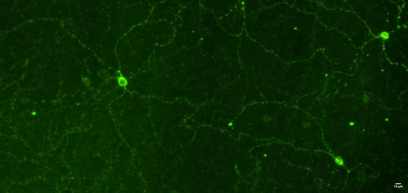
| Applications | ICC |
| Host | Chicken |
| Reactivity | Mouse |
| Note | STRICTLY FOR FURTHER SCIENTIFIC RESEARCH USE ONLY (RUO). MUST NOT TO BE USED IN DIAGNOSTIC OR THERAPEUTIC APPLICATIONS. |
Product Properties
| Clonality | Polyclonal |
| Isotype | IgY |
| Conjugation | Unconjugated |
| Concentration | 200 µg/mL |
| Purification | Affinity-Purified IgY |
| Dilution Range | WB N/AWB Brain N/AIHC N/AICC 1:1000-1:10, 000 |
| Formulation | Phosphate-buffered (10 mM) isotonic (0.9%, w/v) saline (“PBS, pH 7.2) with sodium azide (0.02%, w/v) added as a preservative." |
| Storage Instruction | Store at 4°C in the dark. Under these conditions, the antibodies should have a shelf life of at least twelve months, provided they remain sterile. For longer term storage, aliquot and freeze to avoid freeze-thaw of the antibody. |
Target Information
| Gene Symbol | Opn4 |
| Gene ID | 30044 |
| Uniprot ID | OPN4_MOUSE |
| Immunogen | Synthetic peptide |
| Immunogen Sequence | Mouse |
| Specificity | No cross-reactivity reported |
Additional Info
| Tissue Specificity | Expressed in the retinal pigment epithelium and ganglion cell layer (at protein level). Also expressed in amacrine cell layers of the retina. Weakly expressed in vibrissae, and tail. Isoform 1: Observed with processes in the outer strata of inner plexiform layer (IPL) close to the inner nuclear layer (INL) or is found to be bistratified with processes located both in the inner (ON) or outer (OFF) layers of the IPL (at protein level). A second population of isoform 1 is identified in processes which are confined to the inner layer of the IPL near to the ganglion cell layer (GCL) (at protein level). Isoform 2: About 40 times more abundant than isoform 1 in the retina (at protein level). Isoform 2 is involved in processes localized to the outer IPL or is bistratified with processes in both the inner and outer layers of the IPL (at protein level). Isoform 2 is absent in the processes confined only to the inner layer of the IPL (at protein level). |
| Function | Photoreceptor that binds cis-retinaldehydes. Contributes to pupillar reflex, photoentrainment and other non-image forming responses to light. May be involved in the optokinetic visual tracking response. May be involved in the regulation of retinal hyaloid vessel growth and regression. |
| Protein Name | MelanopsinOpsin-4 |
| Database Links | Reactome: R-MMU-416476Reactome: -MMU-419771 |
| Cellular Localisation | Cell MembraneMulti-Pass Membrane ProteinCell ProjectionAxonDendritePerikaryon |
| Alternative Antibody Names | Anti-Melanopsin antibodyAnti-Opsin-4 antibodyAnti-Opn4 antibodyAnti-Mop antibodyAnti-Mopn antibody |
Information sourced from Uniprot.org