| Host: |
Rabbit |
| Applications: |
WB/IF/IP |
| Reactivity: |
Human/Mouse/Rat |
| Note: |
STRICTLY FOR FURTHER SCIENTIFIC RESEARCH USE ONLY (RUO). MUST NOT TO BE USED IN DIAGNOSTIC OR THERAPEUTIC APPLICATIONS. |
| Short Description: |
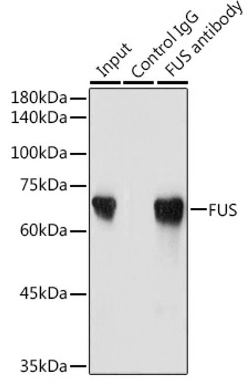
Rabbit polyclonal antibody anti-FUS (297-526) is suitable for use in Western Blot, Immunofluorescence and Immunoprecipitation research applications. |
| Clonality: |
Polyclonal |
| Conjugation: |
Unconjugated |
| Isotype: |
IgG |
| Formulation: |
PBS with 0.02% Sodium Azide, 50% Glycerol, pH7.3. |
| Purification: |
Affinity purification |
| Dilution Range: |
WB 1:500-1:2000IF/ICC 1:50-1:100IP 1:1000-1:5000 |
| Storage Instruction: |
Store at-20°C for up to 1 year from the date of receipt, and avoid repeat freeze-thaw cycles. |
| Gene Symbol: |
FUS |
| Gene ID: |
2521 |
| Uniprot ID: |
FUS_HUMAN |
| Immunogen Region: |
297-526 |
| Immunogen: |
Recombinant fusion protein containing a sequence corresponding to amino acids 297-526 of human FUS (NP_004951.1). |
| Immunogen Sequence: |
TIESVADYFKQIGIIKTNKK TGQPMINLYTDRETGKLKGE ATVSFDDPPSAKAAIDWFDG KEFSGNPIKVSFATRRADFN RGGGNGRGGRGRGGPMGRGG YGGGGSGGGGRGGFPSGGGG GGGQQRAGDWKCPNPTCENM NFSWRNECNQCKAPKPDGPG GGPGGSHMGGNYGDDRRGGR GGYDRGGYRGRGGDRGGFRG GRGGGDRGGFGPGKMDSRGE HRQDRRERPY |
| Tissue Specificity | Ubiquitous. |
| Post Translational Modifications | Arg-216 and Arg-218 are dimethylated, probably to asymmetric dimethylarginine. Phosphorylated in its N-terminal serine residues upon induced DNA damage. ATM and DNA-PK are able to phosphorylate FUS N-terminal region. |
| Function | DNA/RNA-binding protein that plays a role in various cellular processes such as transcription regulation, RNA splicing, RNA transport, DNA repair and damage response. Binds to nascent pre-mRNAs and acts as a molecular mediator between RNA polymerase II and U1 small nuclear ribonucleoprotein thereby coupling transcription and splicing. Binds also its own pre-mRNA and autoregulates its expression.this autoregulation mechanism is mediated by non-sense-mediated decay. Plays a role in DNA repair mechanisms by promoting D-loop formation and homologous recombination during DNA double-strand break repair. In neuronal cells, plays crucial roles in dendritic spine formation and stability, RNA transport, mRNA stability and synaptic homeostasis. |
| Protein Name | Rna-Binding Protein Fus75 Kda Dna-Pairing ProteinOncogene FusOncogene TlsPomp75Translocated In Liposarcoma Protein |
| Database Links | Reactome: R-HSA-72163Reactome: R-HSA-72203 |
| Cellular Localisation | NucleusDisplays A Punctate Pattern Inside The Nucleus And Is Excluded From Nucleoli |
| Alternative Antibody Names | Anti-Rna-Binding Protein Fus antibodyAnti-75 Kda Dna-Pairing Protein antibodyAnti-Oncogene Fus antibodyAnti-Oncogene Tls antibodyAnti-Pomp75 antibodyAnti-Translocated In Liposarcoma Protein antibodyAnti-FUS antibodyAnti-TLS antibody |
Information sourced from Uniprot.org
12 months for antibodies. 6 months for ELISA Kits. Please see website T&Cs for further guidance


![Immunofluorescence analysis of NIH/3T3 cells using [KO Validated] FUS Rabbit polyclonal antibody (STJ27717) at dilution of 1:100. Secondary antibody: Cy3 Goat Anti-Rabbit IgG (H+L) at 1:500 dilution. Blue: DAPI for nuclear staining.](https://cdn11.bigcommerce.com/s-zso2xnchw9/images/stencil/760x760/products/101320/388920/STJ27717_2__45065.1713158496.jpg?c=1)
![Immunofluorescence analysis of C6 cells using [KO Validated] FUS Rabbit polyclonal antibody (STJ27717) at dilution of 1:100. Secondary antibody: Cy3 Goat Anti-Rabbit IgG (H+L) at 1:500 dilution. Blue: DAPI for nuclear staining.](https://cdn11.bigcommerce.com/s-zso2xnchw9/images/stencil/760x760/products/101320/388921/STJ27717_3__73027.1713158496.jpg?c=1)
![Western blot analysis of lysates from wild type (WT) and FUS knockout (KO) 293T cells, using [KO Validated] FUS Rabbit polyclonal antibody (STJ27717) at 1:3000 dilution. Secondary antibody: HRP Goat Anti-Rabbit IgG (H+L) (STJS000856) at 1:10000 dilution. Lysates/proteins: 25 Mu g per lane. Blocking buffer: 3% nonfat dry milk in TBST. Detection: ECL Basic Kit. Exposure time: 1s.](https://cdn11.bigcommerce.com/s-zso2xnchw9/images/stencil/760x760/products/101320/388922/STJ27717_4__93014.1713158497.jpg?c=1)


