

Anti-CD274 antibody [ZR3] {KLH} (STJ180217)
SPECIFICATIONS
ClonalityMonoclonal
HostRabbit
ConjugationKLH
IsotypeIgG
ImmunogenKLH-conjugated linear peptide corresponding tohuman PD-L1
General Information
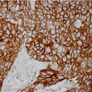
| Short Description | Rabbit monoclonal CD274 antibody for use in IHC-P in human samples. Datasheet included with dilution recommendations, and related reagents. |
| Applications | IHC-P |
| Host | Rabbit |
| Reactivity | Human |
| Note | STRICTLY FOR FURTHER SCIENTIFIC RESEARCH USE ONLY (RUO). MUST NOT TO BE USED IN DIAGNOSTIC OR THERAPEUTIC APPLICATIONS. |
Product Properties
| Clonality | Monoclonal |
| Clone ID | ZR3 |
| Isotype | IgG |
| Conjugation | KLH |
| Purification | Affinity purified |
| Dilution Range | 1:100‐200 |
| Formulation | Tris-HCI buffer containing stabilizing protein (BSA) and <0.1% ProClin |
| Storage Instruction | Store at 2‐8°C for up to 24 months. Predilute: Ready to use, no reconstitution necessary. Concentrate: Use dilution range and appropriate lab‐standardized diluent. Stability after dilution: 7 days at 24°C, 3 months at 2‐8°C, 6months at ‐20°C. |
Target Information
| Gene Symbol | CD274 |
| Gene ID | 29126 |
| Uniprot ID | PD1L1_HUMAN |
| Immunogen | KLH-conjugated linear peptide corresponding tohuman PD-L1 |
| Specificity | Positive control: Placenta or lung adenocarcinoma |
Additional Info
| Tissue Specificity | Highly expressed in the heart, skeletal muscle, placenta and lung. Weakly expressed in the thymus, spleen, kidney and liver. Expressed on activated T- and B-cells, dendritic cells, keratinocytes and monocytes. Isoform 4: Widely expressed, highest in lung, liver and pituitary and in various peripheral blood cells, including neutrophils and some subtypes of lymphoid and myeloid cells. |
| Post Translational Modifications | Ubiquitinated.STUB1 likely mediates polyubiquitination of PD-L1/CD274 triggering its degradation. Ubiquitinated by MARCHF8.leading to degradation. Deubiquitinated by USP22.leading to stabilization. |
| Function | Plays a critical role in induction and maintenance of immune tolerance to self. As a ligand for the inhibitory receptor PDCD1/PD-1, modulates the activation threshold of T-cells and limits T-cell effector response. Through a yet unknown activating receptor, may costimulate T-cell subsets that predominantly produce interleukin-10 (IL10). Can also act as a transcription coactivator: in response to hypoxia, translocates into the nucleus via its interaction with phosphorylated STAT3 and promotes transcription of GSDMC, leading to pyroptosis. The PDCD1-mediated inhibitory pathway is exploited by tumors to attenuate anti-tumor immunity and escape destruction by the immune system, thereby facilitating tumor survival. The interaction with PDCD1/PD-1 inhibits cytotoxic T lymphocytes (CTLs) effector function. The blockage of the PDCD1-mediated pathway results in the reversal of the exhausted T-cell phenotype and the normalization of the anti-tumor response, providing a rationale for cancer immunotherapy. |
| Protein Name | Programmed Cell Death 1 Ligand 1Pd-L1Pdcd1 Ligand 1Programmed Death Ligand 1Hpd-L1B7 Homolog 1B7-H1Cd Antigen Cd274 |
| Database Links | Reactome: R-HSA-389948Reactome: R-HSA-9701898 |
| Cellular Localisation | Cell MembraneSingle-Pass Type I Membrane ProteinEarly Endosome MembraneRecycling Endosome MembraneNucleusAssociates With Cmtm6 At Recycling EndosomesWhere It Is Protected From Being Targeted For Lysosomal DegradationTranslocates To The Nucleus In Response To Hypoxia Via Its Interaction With Phosphorylated Stat3Isoform 1: Cell MembraneIsoform 2: Endomembrane SystemIsoform 4: Secreted |
| Alternative Antibody Names | Anti-Programmed Cell Death 1 Ligand 1 antibodyAnti-Pd-L1 antibodyAnti-Pdcd1 Ligand 1 antibodyAnti-Programmed Death Ligand 1 antibodyAnti-Hpd-L1 antibodyAnti-B7 Homolog 1 antibodyAnti-B7-H1 antibodyAnti-Cd Antigen Cd274 antibodyAnti-CD274 antibodyAnti-B7H1 antibodyAnti-PDCD1L1 antibodyAnti-PDCD1LG1 antibodyAnti-PDL1 antibody |
Information sourced from Uniprot.org