• Western blot analysis of extracts of various cell lines, using HP1 alpha/CBX5rabbit monoclonal antibody (STJ11101944) at 1:1000 dilution. Secondary antibody: HRP Goat Anti-rabbit IgG (H+L) (STJS000856) at 1:10000 dilution. Lysates/proteins: 25 Mu g per lane. Blocking buffer: 3% non-fat dry milk in TBST. Detection: ECL Basic Kit. Exposure time: 10s.
  • Western blot analysis of extracts of various cell lines, using HP1 alpha/CBX5rabbit monoclonal antibody (STJ11101944) at 1:1000 dilution. Secondary antibody: HRP Goat Anti-rabbit IgG (H+L) (STJS000856) at 1:10000 dilution. Lysates/proteins: 25 Mu g per lane. Blocking buffer: 3% non-fat dry milk in TBST. Detection: ECL Basic Kit. Exposure time: 3min.
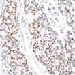
  • Immunohistochemistry analysis of HP1 alpha/CBX5 in paraffin-embedded human colon carcinoma tissue using HP1 alpha/CBX5 rabbit monoclonal antibody (STJ11101944) at a dilution of 1:200 (40x lens). High pressure antigen retrieval was performed with 0. 01 M citrate buffer (pH 6. 0) prior to immunohistochemistry staining.
  • Immunohistochemistry analysis of HP1 alpha/CBX5 in paraffin-embedded human lung cancer tissue using HP1 alpha/CBX5 rabbit monoclonal antibody (STJ11101944) at a dilution of 1:200 (40x lens). High pressure antigen retrieval was performed with 0. 01 M citrate buffer (pH 6. 0) prior to immunohistochemistry staining.
  • Immunohistochemistry analysis of HP1 alpha/CBX5 in paraffin-embedded human thyroid cancer tissue using HP1 alpha/CBX5 rabbit monoclonal antibody (STJ11101944) at a dilution of 1:200 (40x lens). High pressure antigen retrieval was performed with 0. 01 M citrate buffer (pH 6. 0) prior to immunohistochemistry staining.
  • Immunohistochemistry analysis of HP1 alpha/CBX5 in paraffin-embedded mouse brain tissue using HP1 alpha/CBX5 rabbit monoclonal antibody (STJ11101944) at a dilution of 1:200 (40x lens). High pressure antigen retrieval was performed with 0. 01 M citrate buffer (pH 6. 0) prior to immunohistochemistry staining.
  • Immunohistochemistry analysis of HP1 alpha/CBX5 in paraffin-embedded mouse kidney tissue using HP1 alpha/CBX5 rabbit monoclonal antibody (STJ11101944) at a dilution of 1:200 (40x lens). High pressure antigen retrieval was performed with 0. 01 M citrate buffer (pH 6. 0) prior to immunohistochemistry staining.
  • Immunohistochemistry analysis of HP1 alpha/CBX5 in paraffin-embedded mouse lung tissue using HP1 alpha/CBX5 rabbit monoclonal antibody (STJ11101944) at a dilution of 1:200 (40x lens). High pressure antigen retrieval was performed with 0. 01 M citrate buffer (pH 6. 0) prior to immunohistochemistry staining.
  • Immunohistochemistry analysis of HP1 alpha/CBX5 in paraffin-embedded mouse testis tissue using HP1 alpha/CBX5 rabbit monoclonal antibody (STJ11101944) at a dilution of 1:200 (40x lens). High pressure antigen retrieval was performed with 0. 01 M citrate buffer (pH 6. 0) prior to immunohistochemistry staining.
  • Immunohistochemistry analysis of HP1 alpha/CBX5 in paraffin-embedded rat brain tissue using HP1 alpha/CBX5 rabbit monoclonal antibody (STJ11101944) at a dilution of 1:200 (40x lens). High pressure antigen retrieval was performed with 0. 01 M citrate buffer (pH 6. 0) prior to immunohistochemistry staining.
  • Immunohistochemistry analysis of HP1 alpha/CBX5 in paraffin-embedded rat colon tissue using HP1 alpha/CBX5 rabbit monoclonal antibody (STJ11101944) at a dilution of 1:200 (40x lens). High pressure antigen retrieval was performed with 0. 01 M citrate buffer (pH 6. 0) prior to immunohistochemistry staining.
  • Immunofluorescence analysis of C6 cells using HP1 alpha/CBX5rabbit monoclonal antibody (STJ11101944) at dilution of 1:100 (40x lens). Blue: DAPI for nuclear staining.
  • Immunoprecipitation analysis of 300 Mu g extracts of MCF7 cells using 3 Mu g HP1 alpha/CBX5 antibody (STJ11101944). Western blot was performed from the immunoprecipitate using HP1 alpha/CBX5 antibody (STJ11101944) at a dilution of 1:1000.

Anti-CBX5 antibody (92-191) [S4MR] (STJ11101944)

SKU:
STJ11101944

Shipping:
Free Shipping
Current Stock:
Host: Rabbit
Applications: WB/IHC/IF/IP
Reactivity: Human/Mouse/Rat
Note: STRICTLY FOR FURTHER SCIENTIFIC RESEARCH USE ONLY (RUO). MUST NOT TO BE USED IN DIAGNOSTIC OR THERAPEUTIC APPLICATIONS.
Short Description: Rabbit monoclonal antibody anti-HP1 alpha (92-191) is suitable for use in Western Blot, Immunohistochemistry, Immunofluorescence and Immunoprecipitation research applications.
Clonality: Monoclonal
Clone ID: S4MR
Conjugation: Unconjugated
Isotype: IgG
Formulation: PBS with 0.02% Sodium Azide, 0.05% BSA, 50% Glycerol, pH7.3.
Purification: Affinity purification
Dilution Range: WB 1:500-1:1000
IHC-P 1:50-1:200
IF/ICC 1:50-1:200
IP 1:500-1:1000
Storage Instruction: Store at-20°C for up to 1 year from the date of receipt, and avoid repeat freeze-thaw cycles.
Gene Symbol: CBX5
Gene ID: 23468
Uniprot ID: CBX5_HUMAN
Immunogen Region: 92-191
Immunogen: A synthetic peptide corresponding to a sequence within amino acids 92-191 of human HP1 alpha/CBX5 (P45973).
Immunogen Sequence: SNFSNSADDIKSKKKREQSN DIARGFERGLEPEKIIGATD SCGDLMFLMKWKDTDEADLV LAKEANVKCPQIVIAFYEER LTWHAYPEDAENKEKETAKS
Post Translational Modifications Phosphorylation of HP1 and LBR may be responsible for some of the alterations in chromatin organization and nuclear structure which occur at various times during the cell cycle. Phosphorylated during interphase and possibly hyper-phosphorylated during mitosis. Ubiquitinated.
Function Component of heterochromatin that recognizes and binds histone H3 tails methylated at 'Lys-9' (H3K9me), leading to epigenetic repression. In contrast, it is excluded from chromatin when 'Tyr-41' of histone H3 is phosphorylated (H3Y41ph). Can interact with lamin-B receptor (LBR). This interaction can contribute to the association of the heterochromatin with the inner nuclear membrane. Involved in the formation of functional kinetochore through interaction with MIS12 complex proteins.
Protein Name Chromobox Protein Homolog 5
Antigen P25
Heterochromatin Protein 1 Homolog Alpha
Hp1 Alpha
Database Links Reactome: R-HSA-4551638
Reactome: R-HSA-8953750
Reactome: R-HSA-983231
Cellular Localisation Nucleus
Chromosome
Centromere
Colocalizes With Hnrnpu In The Nucleus
Component Of Centromeric And Pericentromeric Heterochromatin
Associates With Chromosomes During Mitosis
Associates Specifically With Chromatin During Metaphase And Anaphase
Localizes To Sites Of Dna Damage
Alternative Antibody Names Anti-Chromobox Protein Homolog 5 antibody
Anti-Antigen P25 antibody
Anti-Heterochromatin Protein 1 Homolog Alpha antibody
Anti-Hp1 Alpha antibody
Anti-CBX5 antibody
Anti-HP1A antibody

Information sourced from Uniprot.org

12 months for antibodies. 6 months for ELISA Kits. Please see website T&Cs for further guidance