-
Immunohistochemical analysis of paraffin-embedded Human stomach. 1, Antibody was diluted at 1:100 (4°C overnight). 2, High-pressure and temperature EDTA, pH8.0 was used for antigen retrieval. 3, Secondary antibody was diluted at 1:200 (room temperature, 30min).
-
Western blot analysis of JK using BRCA1 Polyclonal Antibody diluted at 1:1000. Secondary antibody was diluted at 1:20000
-
Immunohistochemical analysis of paraffin-embedded Human stomach. 1, Antibody was diluted at 1:100 (4°C overnight). 2, High-pressure and temperature EDTA, pH8.0 was used for antigen retrieval. 3, Secondary antibody was diluted at 1:200 (room temperature, 30min).
-
Western blot analysis of various cells using BRCA1 Polyclonal Antibody diluted at 1:1000
-
Western blot analysis of HT29 cells using BRCA1 Polyclonal Antibody diluted at 1:1000
-
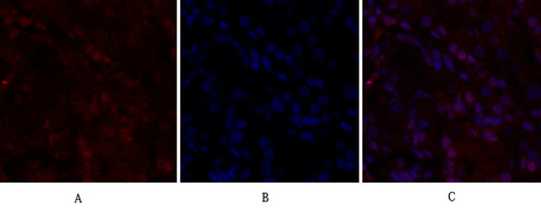
Immunofluorescence analysis of human-stomach tissue. 1, BRCA1 Polyclonal Antibody (red) was diluted at 1:200 (4°C, overnight). 2, Cy3 labled Secondary antibody was diluted at 1:300 (room temperature, 50min).3, Picture B: DAPI (blue) 10min. Picture A:Target. Picture B: DAPI. Picture C: merge of A+B
-
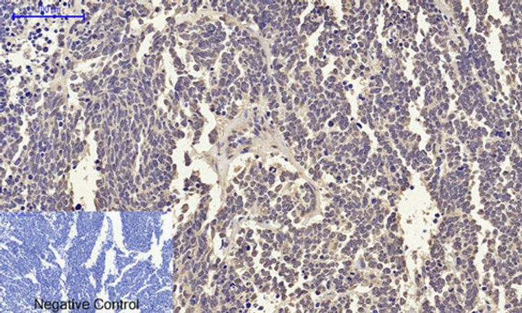
Immunohistochemical analysis of paraffin-embedded Rat-lung tissue. 1, BRCA1 Polyclonal Antibody was diluted at 1:200 (4°C, overnight). 2, Sodium citrate pH 6.0 was used for antibody retrieval (>98°C, 20min). 3, Secondary antibody was diluted at 1:200 (room tempeRature, 30min). Negative control was used by secondary antibody only.
-
Immunohistochemical analysis of paraffin-embedded Human-uterus-cancer tissue. 1, BRCA1 Polyclonal Antibody was diluted at 1:200 (4°C, overnight). 2, Sodium citrate pH 6.0 was used for antibody retrieval (>98°C, 20min). 3, Secondary antibody was diluted at 1:200 (room tempeRature, 30min). Negative control was used by secondary antibody only.
-
Immunohistochemical analysis of paraffin-embedded Human-Tonsil tissue. 1, BRCA1 Polyclonal Antibody was diluted at 1:200 (4°C, overnight). 2, Sodium citrate pH 6.0 was used for antibody retrieval (>98°C, 20min). 3, Secondary antibody was diluted at 1:200 (room tempeRature, 30min). Negative control was used by secondary antibody only.
-
Western blot analysis of various cells using primary antibody diluted at 1:1000 (4°C overnight). Secondary antibody:Goat Anti-rabbit IgG IRDye 800 ( diluted at 1:5000, 25°C, 1 hour). Cell lysate was extracted by Minute Plasma Membrane Protein Isolation and Cell Fractionation Kit (SM-005, Inventbiotech, MN, USA).
-
Immunohistochemical analysis of paraffin-embedded Human-uterus tissue. 1, BRCA1 Polyclonal Antibody was diluted at 1:200 (4°C, overnight). 2, Sodium citrate pH 6.0 was used for antibody retrieval (>98°C, 20min). 3, Secondary antibody was diluted at 1:200 (room tempeRature, 30min). Negative control was used by secondary antibody only.
-
Immunofluorescence analysis of rat-lung tissue. 1, BRCA1 Polyclonal Antibody (red) was diluted at 1:200 (4°C, overnight). 2, Cy3 labled Secondary antibody was diluted at 1:300 (room temperature, 50min).3, Picture B: DAPI (blue) 10min. Picture A:Target. Picture B: DAPI. Picture C: merge of A+B
-
Immunofluorescence analysis of rat-lung tissue. 1, BRCA1 Polyclonal Antibody (red) was diluted at 1:200 (4°C, overnight). 2, Cy3 labled Secondary antibody was diluted at 1:300 (room temperature, 50min).3, Picture B: DAPI (blue) 10min. Picture A:Target. Picture B: DAPI. Picture C: merge of A+B
-
Immunofluorescence analysis of human-stomach tissue. 1, BRCA1 Polyclonal Antibody (red) was diluted at 1:200 (4°C, overnight). 2, Cy3 labled Secondary antibody was diluted at 1:300 (room temperature, 50min).3, Picture B: DAPI (blue) 10min. Picture A:Target. Picture B: DAPI. Picture C: merge of A+B
-
Immunohistochemical analysis of paraffin-embedded Human stomach. 1, Antibody was diluted at 1:100 (4°C overnight). 2, High-pressure and temperature EDTA, pH8.0 was used for antigen retrieval. 3, Secondary antibody was diluted at 1:200 (room temperature, 30min).
-
Immunohistochemical analysis of paraffin-embedded Human-lung-cancer tissue. 1, BRCA1 Polyclonal Antibody was diluted at 1:200 (4°C, overnight). 2, Sodium citrate pH 6.0 was used for antibody retrieval (>98°C, 20min). 3, Secondary antibody was diluted at 1:200 (room tempeRature, 30min). Negative control was used by secondary antibody only.
-
Immunohistochemistry analysis of paraffin-embedded human breast carcinoma tissue, using BRCA1 Antibody. The picture on the right is blocked with the synthesized peptide.
-
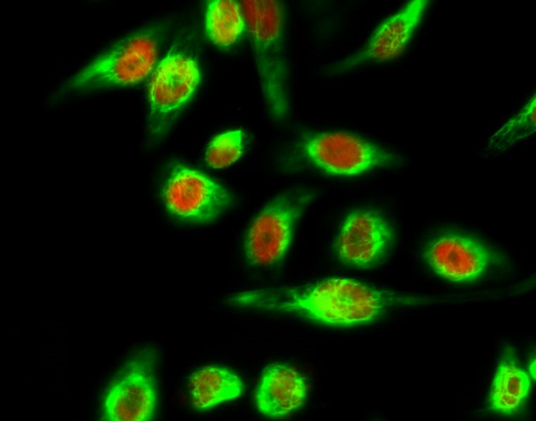
Immunofluorescence analysis of Hela cell. 1, BRCA1 Polyclonal Antibody (green) was diluted at 1:200 (4°C overnight). (red) was diluted at 1:200 (4°C overnight). 2, Goat Anti Rabbit Alexa Fluor 488 Catalog: (NA was diluted at 1:1000 (room temperature, 50min). Goat Anti Mouse Alexa Fluor 594 Catalog: (NA was diluted at 1:1000 (room temperature, 50min).


