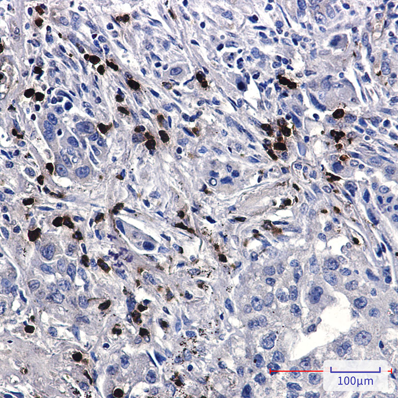
| Tissue Specificity | Highly expressed in brain.strongly reduced in post-mortem elderly subjects with Alzheimer disease. Isoform 4: Expressed preferentially in the brain. |
| Post Translational Modifications | Phosphorylation at Ser-610 by SGK1 promotes its localization to the nucleus. Phosphorylated following nuclear translocation. Phosphorylation at Tyr-547 by ABL1 enhances transcriptional activation activity and reduces the affinity for RASD1/DEXRAS1. Phosphorylated at Ser-459 by PKC upon insulin activation. Acetylation at Lys-204 and Lys-701 by KAT5 promotes its transcription activator activity. Polyubiquitination by RNF157 leads to degradation by the proteasome. |
| Function | Transcription coregulator that can have both coactivator and corepressor functions. Adapter protein that forms a transcriptionally active complex with the gamma-secretase-derived amyloid precursor protein (APP) intracellular domain. Plays a central role in the response to DNA damage by translocating to the nucleus and inducing apoptosis. May act by specifically recognizing and binding histone H2AX phosphorylated on 'Tyr-142' (H2AXY142ph) at double-strand breaks (DSBs), recruiting other pro-apoptosis factors such as MAPK8/JNK1. Required for histone H4 acetylation at double-strand breaks (DSBs). Its ability to specifically bind modified histones and chromatin modifying enzymes such as KAT5/TIP60, probably explains its transcription activation activity. Functions in association with TSHZ3, SET and HDAC factors as a transcriptional repressor, that inhibits the expression of CASP4. Associates with chromatin in a region surrounding the CASP4 transcriptional start site(s). Involved in hippocampal neurite branching and neuromuscular junction formation, as a result plays a role in spatial memory functioning. Plays a role in the maintenance of lens transparency. May play a role in muscle cell strength. Acts as a molecular adapter that functions in neurite outgrowth by activating the RAC1-ARF6 axis upon insulin treatment. |
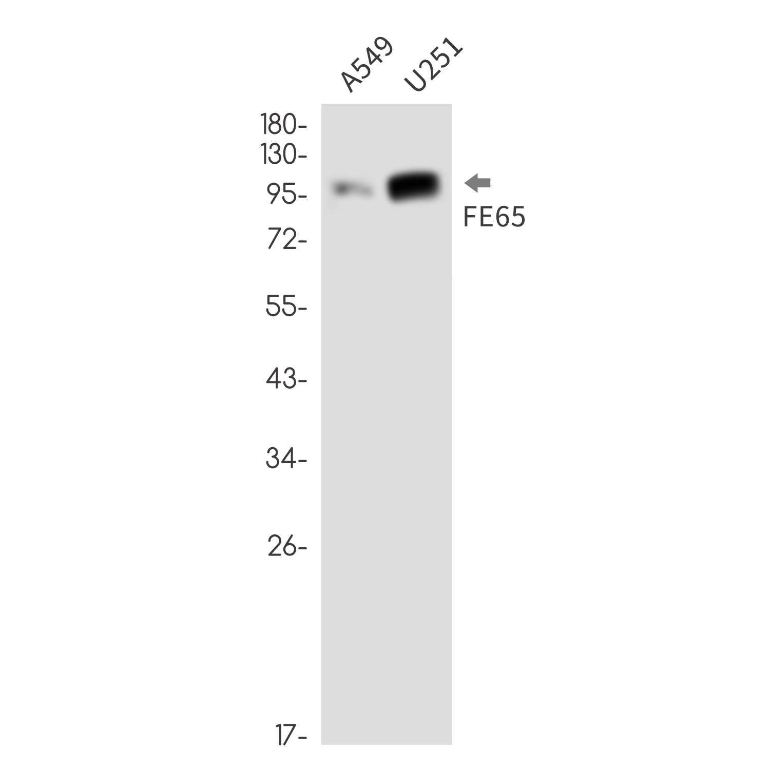
| Protein Name | Amyloid Beta Precursor Protein Binding Family B Member 1Amyloid-Beta A4 Precursor Protein-Binding Family B Member 1Protein Fe65 |
| Database Links | Reactome: R-HSA-5693565 |
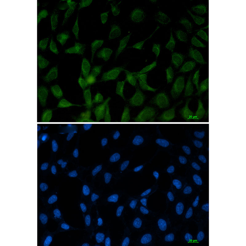
| Cellular Localisation | Cell MembraneCytoplasmNucleusCell ProjectionGrowth ConeNucleus SpeckleColocalizes With Tshz3 In Axonal Growth ConeColocalizes With Tshz3 In The NucleusIn Normal ConditionsIt Mainly Localizes To The CytoplasmWhile A Small Fraction Is Tethered To The Cell Membrane Via Its Interaction With AppFollowing Exposure To Dna Damaging AgentsIt Is Released From Cell Membrane And Translocates To The NucleusNuclear Translocation Is Under The Regulation Of AppColocalizes With Nek6 At The Nuclear SpecklesPhosphorylation At Ser-610 By Sgk1 Promotes Its Localization To The Nucleus |
| Alternative Antibody Names | Anti-Amyloid Beta Precursor Protein Binding Family B Member 1 antibodyAnti-Amyloid-Beta A4 Precursor Protein-Binding Family B Member 1 antibodyAnti-Protein Fe65 antibodyAnti-APBB1 antibodyAnti-FE65 antibodyAnti-RIR antibody |
Information sourced from Uniprot.org


