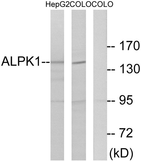
• Western blot analysis of lysates from COLO and HepG2 cells, using ALPK1 Antibody. The lane on the right is blocked with the synthesized peptide.
  • Western blot analysis of the lysates from 293 cells using ALPK1 antibody.
  • Western blot analysis of various cells using Alpha-protein Kinase 1 Polyclonal Antibody. Secondary antibody was diluted at 1:20000

Anti-ALPK1 antibody (11-60 aa) (STJ96366)

SKU:
STJ96366

Current Stock:
Host: Rabbit
Applications: WB/IHC/IF/ELISA
Reactivity: Human/Mouse
Note: STRICTLY FOR FURTHER SCIENTIFIC RESEARCH USE ONLY (RUO). MUST NOT TO BE USED IN DIAGNOSTIC OR THERAPEUTIC APPLICATIONS.
Short Description: Rabbit polyclonal antibody anti-Alpha-protein kinase 1 (11-60 aa) is suitable for use in Western Blot, Immunohistochemistry, Immunofluorescence and ELISA research applications.
Clonality: Polyclonal
Conjugation: Unconjugated
Isotype: IgG
Formulation: Liquid in PBS containing 50% Glycerol, 0.5% BSA and 0.02% Sodium Azide.
Purification: The antibody was affinity-purified from rabbit antiserum by affinity-chromatography using epitope-specific immunogen.
Concentration: 1 mg/mL
Dilution Range: WB 1:500-1:2000
IHC 1:100-1:300
IF 1:200-1:1000
ELISA 1:20000
Storage Instruction: Store at-20°C for up to 1 year from the date of receipt, and avoid repeat freeze-thaw cycles.
Gene Symbol: ALPK1
Gene ID: 80216
Uniprot ID: ALPK1_HUMAN
Immunogen Region: 11-60 aa
Specificity: Alpha-protein Kinase 1 Polyclonal Antibody detects endogenous levels of Alpha-protein Kinase 1 protein.
Immunogen: The antiserum was produced against synthesized peptide derived from the human ALPK1 at the amino acid range 11-60
Function Serine/threonine-protein kinase that detects bacterial pathogen-associated molecular pattern metabolites (PAMPs) and initiates an innate immune response, a critical step for pathogen elimination and engagement of adaptive immunity. Specifically recognizes and binds ADP-D-glycero-beta-D-manno-heptose (ADP-Heptose), a potent PAMP present in all Gram-negative and some Gram-positive bacteria. ADP-Heptose-binding stimulates its kinase activity to phosphorylate and activate TIFA, triggering pro-inflammatory NF-kappa-B signaling. May be involved in monosodium urate monohydrate (MSU)-induced inflammation by mediating phosphorylation of unconventional myosin MYO9A. May also play a role in apical protein transport by mediating phosphorylation of unconventional myosin MYO1A. May play a role in ciliogenesis.
Protein Name Alpha-Protein Kinase 1
Chromosome 4 Kinase
Lymphocyte Alpha-Protein Kinase
Database Links Reactome: R-HSA-445989
Reactome: R-HSA-9645460
Cellular Localisation Cytoplasm
Cytosol
Cytoskeleton
Spindle Pole
Microtubule Organizing Center
Centrosome
Cell Projection
Cilium
Localized At The Base Of Primary Cilia
Alternative Antibody Names Anti-Alpha-Protein Kinase 1 antibody
Anti-Chromosome 4 Kinase antibody
Anti-Lymphocyte Alpha-Protein Kinase antibody
Anti-ALPK1 antibody
Anti-KIAA1527 antibody
Anti-LAK antibody

Information sourced from Uniprot.org

12 months for antibodies. 6 months for ELISA Kits. Please see website T&Cs for further guidance