
Nanobodies (VHH Single Domain Antibodies)
Nanobodies in our range are recombinant VHH domains derived from the heavy-chain-only antibodies found in camelid serum, produced in alpaca. Each nanobody is a single binding domain of approximately 15 kDa, roughly one-tenth the size of a conventional IgG, and binds antigen without pairing with a light chain.
Browse All NanobodiesNanobody vs Antibody: Key Differences
A nanobody and a conventional IgG antibody differ in more than size. Select a row for the research context behind each property.
Molecular weight ~15 kDa ~150 kDa ▶
At approximately 15 kDa, a nanobody is roughly one-tenth the molecular weight of a conventional IgG. This small size lets nanobodies diffuse rapidly through tissue and access densely packed cellular environments, which is part of why they are well suited to super-resolution microscopy techniques such as STORM and STED, where the 10 to 15 nm linkage error of an IgG-secondary antibody system can limit spatial resolution.
Structure Single VHH domain Two heavy + two light chains ▶
A nanobody consists of a single VHH domain with three complementarity-determining regions. The CDR3 loop is frequently longer than the equivalent loop in a conventional VH domain, which lets it reach into enzyme clefts and other recessed sites. A conventional IgG instead relies on a paired heavy and light chain to form its antigen-binding site.
Cryptic epitope access Yes Limited ▶
Nanobodies are the preferred choice where the epitope of interest sits within an enzyme active site or a conformationally restricted receptor pocket that is sterically inaccessible to a full-size IgG. This also makes them useful in high-resolution imaging settings where secondary antibody background is a problem.
Thermal stability High (reversible unfolding) Lower (irreversible denaturation) ▶
The disulfide-stabilised core of a VHH domain gives it stability characteristics distinct from a full-length antibody. This favours nanobodies for competitive assay formats or protocols that involve harsher fixation or extended incubation at elevated temperature.
Production Recombinant (E. coli, yeast) Hybridoma or recombinant ▶
All nanobodies in our range are produced recombinantly, which removes the batch-to-batch variability associated with hybridoma-derived antibodies and gives a consistent, traceable reagent for repeat experiments. Recombinant production also makes it straightforward to engineer Fc fusions, enzyme conjugates or fluorescent protein fusions.
Live-cell imaging Well-suited (small, stable) Challenging at full IgG size ▶
A nanobody's size and stability under live-cell conditions support real-time protein tracking, including as the antigen-binding half of a genetically encoded chromobody fusion expressed inside the cell itself. See our fluorescent protein nanobodies below.
Engineering Straightforward (single domain) More complex ▶
Because a nanobody is a single genetic construct rather than a paired heavy and light chain, it is comparatively simple to fuse to an Fc region, an enzyme or a fluorescent protein. See our Chimeric Antibodies range for nanobody-Fc fusion formats.
CD Marker Nanobodies
CD8, CD7 and CD19 are among the most frequently ordered clones in our nanobody range, used as core lineage markers across T-cell and B-cell immunology.

Anti-CD8 Nanobody [SAA2076] (STJN000581)
CD8 marks cytotoxic T lymphocytes and pairs with the T-cell receptor to recognise MHC class I-presented antigen. This clone is validated for ELISA against human CD8 alpha chain.
View product →
Anti-CD7 Nanobody [SAA1260] (STJN000127)
CD7 is expressed on T cells and NK cells from early development onward and is a common marker in T-cell leukaemia panels. Validated for ELISA and flow cytometry against human CD7.
View product →
Anti-CD19 Nanobody [SAA1246] (STJN000155)
CD19 is a pan-B-cell marker and coreceptor for the B-cell antigen receptor, and the most established CAR-T target in B-cell malignancies. Validated for ELISA and flow cytometry against human CD19.
View product →Fluorescent Protein Nanobodies
Nanobodies against fluorescent proteins are used to track tagged proteins inside living cells, either as direct probes or as the antigen-binding half of a genetically encoded "chromobody" fusion expressed inside the cell itself. Their small size and stable folding let them enter densely packed intracellular compartments that are difficult for a full IgG-secondary antibody pair to reach.
Epitope Tag Nanobodies
Nanobodies raised against epitope tags give researchers a compact affinity reagent for detecting or purifying a tagged construct without a second, species-matched secondary antibody. The V5 nanobody below is validated for ELISA and immunofluorescence; the mCherry nanobody is currently validated for ELISA.
Anti-VHH Nanobodies for Nanobody Characterisation
An anti-VHH reagent recognises the shared framework of a camelid VHH domain rather than a specific antigen-binding loop, so a single anti-VHH nanobody can detect a wide range of unrelated nanobody constructs. This is used to confirm expression of a newly generated VHH construct and to serve as an isotype control in flow cytometry assays built around a VHH-based binder.
Anti-GFP Nanobody [SAA0958]
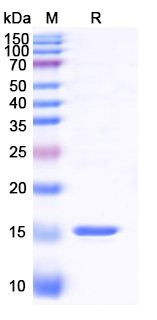
One of our most requested anti-GFP clones, shown here with both its immunofluorescence and SDS-PAGE validation data alongside the full product specification.
Anti-GFP Nanobody [SAA0958] (STJN000449)
Validated by immunofluorescence in GFP-transfected cells, with signal confirmed against a DAPI nuclear counterstain, and by SDS-PAGE for purity ahead of release. A His-tagged format allows detection through a standard anti-His secondary step.
View product →
Where Nanobodies Are Used
Immunofluorescence
Reduced linkage error versus primary plus secondary antibody systems improves spatial resolution in fixed and live-cell imaging.
Western Blot
Clean, low-background detection of nanobody-bound targets, with the small size supporting direct anti-VHH secondary detection.
Flow Cytometry
Reduced steric hindrance on cell surfaces allows access to densely expressed markers such as CD8, CD7 and CD19.
Immunoprecipitation
Nanobody resin is a widely adopted tool for rapid, gentle pulldown of tagged fusion proteins from lysate, as with GFP nanobody resin.1
ELISA
Thermal and incubation stability supports sandwich ELISA formats, as both capture and detection reagent.
Looking for a Specific Nanobody?
Browse the full range of over 700 VHH single domain antibodies, including our complete set of anti-GFP nanobody clones.
Browse All Nanobodies1. Rothbauer U, Zolghadr K, Muyldermans S, Schepers A, Cardoso MC, Leonhardt H. A versatile nanotrap for biochemical and functional studies with fluorescent fusion proteins. Mol Cell Proteomics. 2008;7(2):282-289.