

. STJ95612")

Anti-SEPTIN2 antibody (103-152 aa) (STJ95612)
SPECIFICATIONS
ClonalityPolyclonal
HostRabbit
ConjugationUnconjugated
IsotypeIgG
ImmunogenThe antiserum was produced against synthesized peptide derived from the human SEPT2 at the amino acid range 103-152
General Information
| Short Description | Rabbit polyclonal anti-Septin-2 (103-152 aa) for use in WB and IHC in Human, Mouse and Rat samples. Datasheet included with dilution recommendations, and related reagents. |
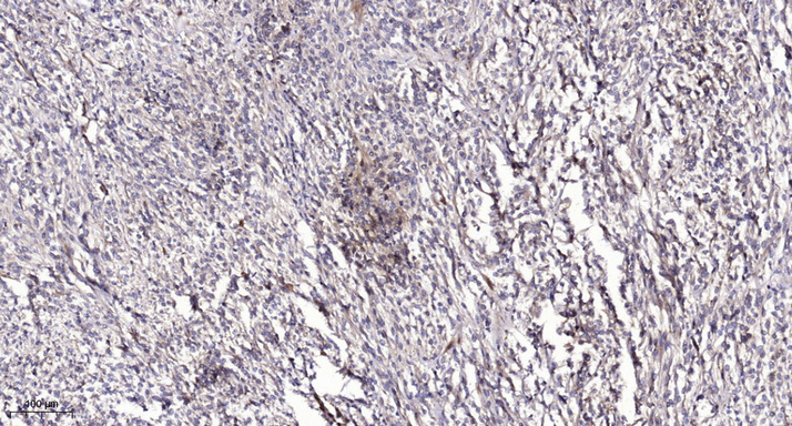
| Applications | WB/IHC |
| Host | Rabbit |
| Reactivity | Human/Mouse/Rat |
| Note | STRICTLY FOR FURTHER SCIENTIFIC RESEARCH USE ONLY (RUO). MUST NOT TO BE USED IN DIAGNOSTIC OR THERAPEUTIC APPLICATIONS. |
Product Properties
| Clonality | Polyclonal |
| Isotype | IgG |
| Conjugation | Unconjugated |
| Concentration | 1 mg/mL |
| Purification | The antibody was affinity-purified from rabbit antiserum by affinity-chromatography using epitope-specific immunogen. |
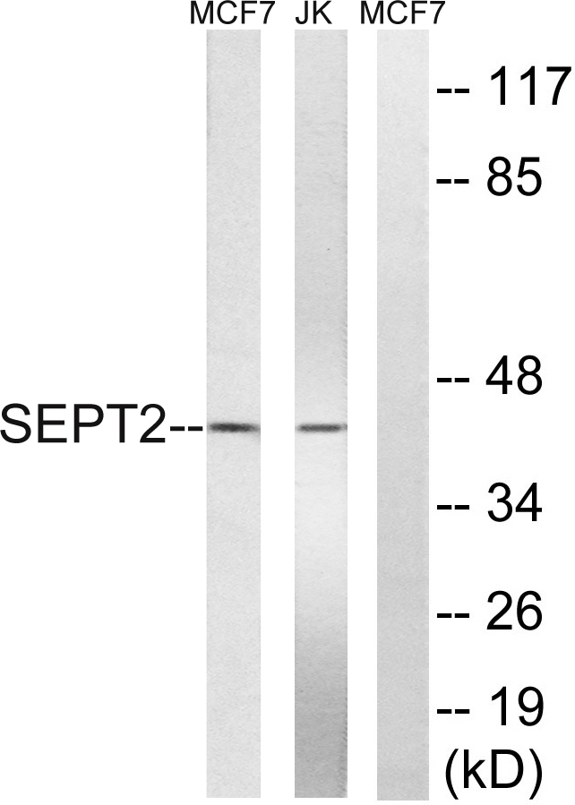
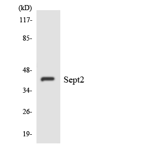
| Dilution Range | WB 1:500-2000IHC-P 1:50-300 |
| Formulation | Liquid in PBS containing 50% Glycerol, 0.5% BSA and 0.02% Sodium Azide. |
| Storage Instruction | Store at-20°C for up to 1 year from the date of receipt, and avoid repeat freeze-thaw cycles. |
Target Information
| Gene Symbol | SEPTIN2 |
| Gene ID | 4735 |
| Uniprot ID | SEPT2_HUMAN |
| Immunogen | The antiserum was produced against synthesized peptide derived from the human SEPT2 at the amino acid range 103-152 |
| Immunogen Region | 103-152 aa |
| Specificity | Septin 2 Polyclonal Antibody detects endogenous levels of Septin 2 protein. |
Additional Info
| Function | Filament-forming cytoskeletal GTPase. Forms a filamentous structure with SEPTIN12, SEPTIN6, SEPTIN2 and probably SEPTIN4 at the sperm annulus which is required for the structural integrity and motility of the sperm tail during postmeiotic differentiation. Required for normal organization of the actin cytoskeleton. Plays a role in the biogenesis of polarized columnar-shaped epithelium by maintaining polyglutamylated microtubules, thus facilitating efficient vesicle transport, and by impeding MAP4 binding to tubulin. Required for the progression through mitosis. Forms a scaffold at the midplane of the mitotic splindle required to maintain CENPE localization at kinetochores and consequently chromosome congression. During anaphase, may be required for chromosome segregation and spindle elongation. Plays a role in ciliogenesis and collective cell movements. In cilia, required for the integrity of the diffusion barrier at the base of the primary cilium that prevents diffusion of transmembrane proteins between the cilia and plasma membranes: probably acts by regulating the assembly of the tectonic-like complex (also named B9 complex) by localizing TMEM231 protein. May play a role in the internalization of 2 intracellular microbial pathogens, Listeria monocytogenes and Shigella flexneri. |
| Protein Name | Septin-2Neural Precursor Cell Expressed Developmentally Down-Regulated Protein 5Nedd-5 |
| Database Links | Reactome: R-HSA-5620912 |
| Cellular Localisation | CytoplasmCytoskeletonSpindleChromosomeCentromereKinetochoreCleavage FurrowMidbodyCell CortexCell ProjectionCilium MembraneCiliumFlagellumIn Metaphase CellsLocalized Within The Microtubule SpindleAt The Metaphase PlateIn Close Apposition To The Kinetochores Of The Congressed ChromosomesIn Cells Undergoing CytokinesisLocalized To The MidbodyThe Ingressing Cleavage FurrowAnd The Central SpindleDuring Bacterial InfectionDisplays A Collar Shape Structure Next To Actin At The Pole Of Invading BacteriaIn Epithelial CellsColocalizes With Polyglutamylated Tubulin Around The Trans-Golgi NetworkAs Well As Juxatnuclear And Proximal Golgi ApparatusLocalizes At The Base Of The Cilia Near The Morphological Distinction Between The Cilia And Plasma MembranesFound In The Sperm Annulus |
| Alternative Antibody Names | Anti-Septin-2 antibodyAnti-Neural Precursor Cell Expressed Developmentally Down-Regulated Protein 5 antibodyAnti-Nedd-5 antibodyAnti-SEPTIN2 antibodyAnti-DIFF6 antibodyAnti-KIAA0158 antibodyAnti-NEDD5 antibodyAnti-SEPT2 antibody |
Information sourced from Uniprot.org