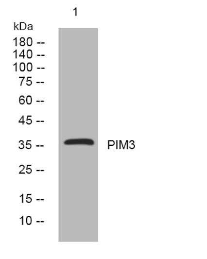
| Tissue Specificity | Detected in various tissues, including the heart, brain, lung, kidney, spleen, placenta, skeletal muscle, and peripheral blood leukocytes. Not found or barely expressed in the normal adult endoderm-derived organs such as colon, thymus, liver, or small intestine. However, expression is augmented in premalignant and malignant lesions of these organs. |
| Post Translational Modifications | Ubiquitinated, leading to proteasomal degradation. Phosphorylated. Interaction with PPP2CA promotes dephosphorylation. |
| Function | Proto-oncogene with serine/threonine kinase activity that can prevent apoptosis, promote cell survival and protein translation. May contribute to tumorigenesis through: the delivery of survival signaling through phosphorylation of BAD which induces release of the anti-apoptotic protein Bcl-X(L), the regulation of cell cycle progression, protein synthesis and by regulation of MYC transcriptional activity. Additionally to this role on tumorigenesis, can also negatively regulate insulin secretion by inhibiting the activation of MAPK1/3 (ERK1/2), through SOCS6. Involved also in the control of energy metabolism and regulation of AMPK activity in modulating MYC and PPARGC1A protein levels and cell growth. |
| Protein Name | Serine/Threonine-Protein Kinase Pim-3 |
| Cellular Localisation | Cytoplasm |
| Alternative Antibody Names | Anti-Serine/Threonine-Protein Kinase Pim-3 antibodyAnti-PIM3 antibody |
Information sourced from Uniprot.org


