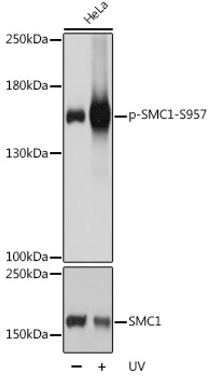
| Host: |
Rabbit |
| Applications: |
WB/IHC/IP |
| Reactivity: |
Human/Mouse/Rat |
| Note: |
STRICTLY FOR FURTHER SCIENTIFIC RESEARCH USE ONLY (RUO). MUST NOT TO BE USED IN DIAGNOSTIC OR THERAPEUTIC APPLICATIONS. |
| Short Description: |
Rabbit polyclonal antibody anti-Phospho-SMC1A-S957 is suitable for use in Western Blot, Immunohistochemistry and Immunoprecipitation research applications. |
| Clonality: |
Polyclonal |
| Conjugation: |
Unconjugated |
| Isotype: |
IgG |
| Formulation: |
PBS with 0.02% Sodium Azide, 50% Glycerol, pH7.3. |
| Purification: |
Affinity purification |
| Dilution Range: |
WB 1:500-1:2000IHC-P 1:50-1:200IP 1:500-1:1000 |
| Storage Instruction: |
Store at-20°C for up to 1 year from the date of receipt, and avoid repeat freeze-thaw cycles. |
| Gene Symbol: |
SMC1A |
| Gene ID: |
8243 |
| Uniprot ID: |
SMC1A_HUMAN |
| Immunogen: |
A synthetic phosphorylated peptide around S957 of human SMC1A (NP_006297.2). |
| Immunogen Sequence: |
GSSQG |
| Post Translational Modifications | Ubiquitinated by the DCX(DCAF15) complex, leading to its degradation. Phosphorylated by ATM upon ionizing radiation in a NBS1-dependent manner. Phosphorylated by ATR upon DNA methylation in a MSH2/MSH6-dependent manner. Phosphorylation of Ser-957 and Ser-966 activates it and is required for S-phase checkpoint activation. |
| Function | Involved in chromosome cohesion during cell cycle and in DNA repair. Central component of cohesin complex. The cohesin complex is required for the cohesion of sister chromatids after DNA replication. The cohesin complex apparently forms a large proteinaceous ring within which sister chromatids can be trapped. At anaphase, the complex is cleaved and dissociates from chromatin, allowing sister chromatids to segregate. The cohesin complex may also play a role in spindle pole assembly during mitosis. Involved in DNA repair via its interaction with BRCA1 and its related phosphorylation by ATM, or via its phosphorylation by ATR. Works as a downstream effector both in the ATM/NBS1 branch and in the ATR/MSH2 branch of S-phase checkpoint. |
| Protein Name | Structural Maintenance Of Chromosomes Protein 1aSmc Protein 1aSmc-1-AlphaSmc-1aSb1.8 |
| Database Links | Reactome: R-HSA-1221632Reactome: R-HSA-2467813Reactome: R-HSA-2468052Reactome: R-HSA-2470946Reactome: R-HSA-2500257Reactome: R-HSA-3108214Reactome: R-HSA-9018519 |
| Cellular Localisation | NucleusChromosomeCentromereKinetochoreAssociates With ChromatinBefore Prophase It Is Scattered Along Chromosome ArmsDuring ProphaseMost Of Cohesin Complexes Dissociate From Chromatin Probably Because Of Phosphorylation By PlkExcept At CentromeresWhere Cohesin Complexes RemainAt AnaphaseThe Rad21 Subunit Of The Cohesin Complex Is CleavedLeading To The Dissociation Of The Complex From ChromosomesAllowing Chromosome SeparationIn Germ CellsCohesin Complex Dissociates From Chromatin At Prophase IAnd May Be Replaced By A Meiosis-Specific Cohesin ComplexThe Phosphorylated Form On Ser-957 And Ser-966 Associates With Chromatin During G1/S/G2 Phases But Not During M PhaseSuggesting That Phosphorylation Does Not Regulate Cohesin FunctionIntegral Component Of The Functional Centromere-Kinetochore Complex At The Kinetochore Region During Mitosis |
| Alternative Antibody Names | Anti-Structural Maintenance Of Chromosomes Protein 1a antibodyAnti-Smc Protein 1a antibodyAnti-Smc-1-Alpha antibodyAnti-Smc-1a antibodyAnti-Sb1.8 antibodyAnti-SMC1A antibodyAnti-DXS423E antibodyAnti-KIAA0178 antibodyAnti-SB1.8 antibodyAnti-SMC1 antibodyAnti-SMC1L1 antibody |
Information sourced from Uniprot.org
12 months for antibodies. 6 months for ELISA Kits. Please see website T&Cs for further guidance


