


. STJ92992")
. 2, Goat Anti Rabbit STJ92992")

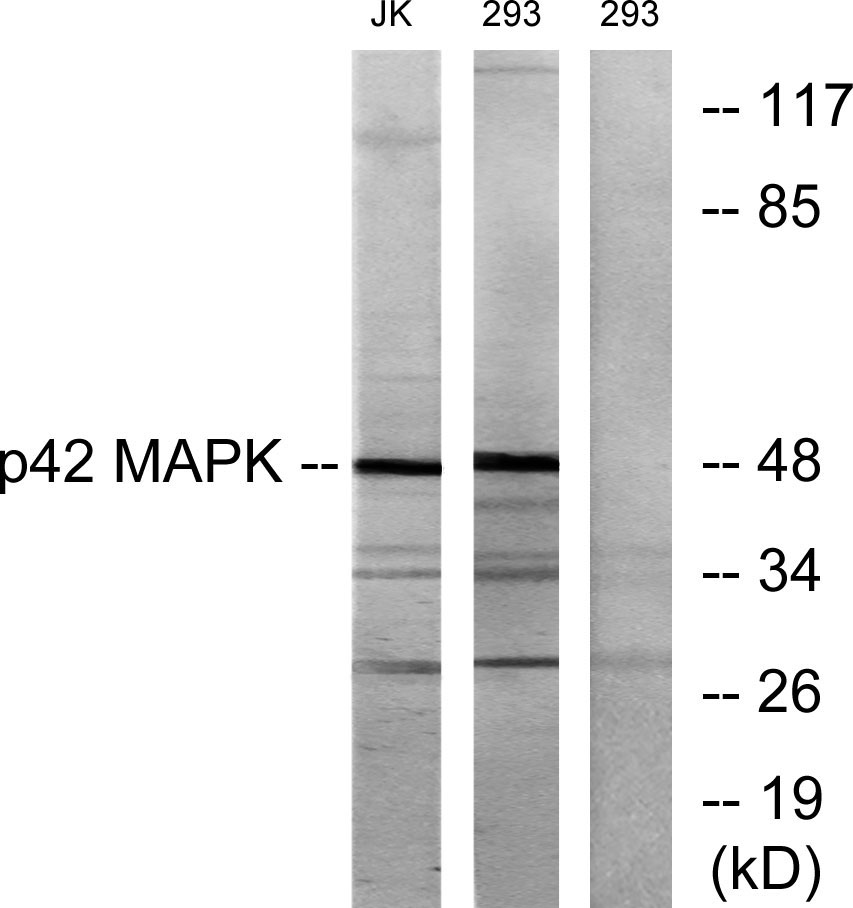
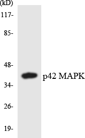
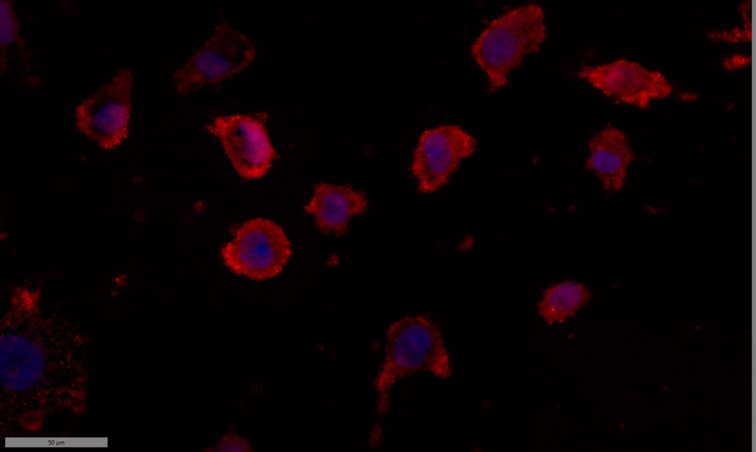
Anti-MAPK1 antibody (136-185 aa) (STJ92992)
SPECIFICATIONS
ClonalityPolyclonal
HostRabbit
ConjugationUnconjugated
IsotypeIgG
ImmunogenThe antiserum was produced against synthesized peptide derived from the human p42 MAPK at the amino acid range 136-185
General Information
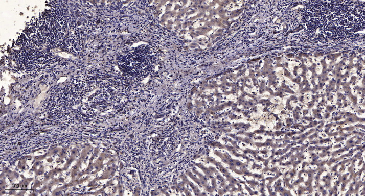
| Short Description | Rabbit polyclonal anti-Mitogen-activated protein kinase 1 (136-185 aa) for use in WB, IHC, IF and ELISA in Human, Mouse and Rat samples. Datasheet included with dilution recommendations, and related reagents. |
| Applications | WB/IHC/IF/ELISA |
| Host | Rabbit |
| Reactivity | Human/Mouse/Rat |
| Note | STRICTLY FOR FURTHER SCIENTIFIC RESEARCH USE ONLY (RUO). MUST NOT TO BE USED IN DIAGNOSTIC OR THERAPEUTIC APPLICATIONS. |
Product Properties
| Clonality | Polyclonal |
| Isotype | IgG |
| Conjugation | Unconjugated |
| Concentration | 1 mg/mL |
| Purification | The antibody was affinity-purified from rabbit antiserum by affinity-chromatography using epitope-specific immunogen. |
| Dilution Range | WB 1:500-1:2000IHC 1:100-1:300IF 1:200-1:1000ELISA 1:5000 |
| Formulation | Liquid in PBS containing 50% Glycerol, 0.5% BSA and 0.02% Sodium Azide. |
| Storage Instruction | Store at-20°C for up to 1 year from the date of receipt, and avoid repeat freeze-thaw cycles. |
Target Information
| Gene Symbol | MAPK1 |
| Gene ID | 5594 |
| Uniprot ID | MK01_HUMAN |
| Immunogen | The antiserum was produced against synthesized peptide derived from the human p42 MAPK at the amino acid range 136-185 |
| Immunogen Region | 136-185 aa |
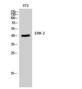
| Specificity | ERK 2 Polyclonal Antibody detects endogenous levels of ERK 2 protein. |
Additional Info
| Function | Serine/threonine kinase which acts as an essential component of the MAP kinase signal transduction pathway. MAPK1/ERK2 and MAPK3/ERK1 are the 2 MAPKs which play an important role in the MAPK/ERK cascade. They participate also in a signaling cascade initiated by activated KIT and KITLG/SCF. Depending on the cellular context, the MAPK/ERK cascade mediates diverse biological functions such as cell growth, adhesion, survival and differentiation through the regulation of transcription, translation, cytoskeletal rearrangements. The MAPK/ERK cascade also plays a role in initiation and regulation of meiosis, mitosis, and postmitotic functions in differentiated cells by phosphorylating a number of transcription factors. About 160 substrates have already been discovered for ERKs. Many of these substrates are localized in the nucleus, and seem to participate in the regulation of transcription upon stimulation. However, other substrates are found in the cytosol as well as in other cellular organelles, and those are responsible for processes such as translation, mitosis and apoptosis. Moreover, the MAPK/ERK cascade is also involved in the regulation of the endosomal dynamics, including lysosome processing and endosome cycling through the perinuclear recycling compartment (PNRC).as well as in the fragmentation of the Golgi apparatus during mitosis. The substrates include transcription factors (such as ATF2, BCL6, ELK1, ERF, FOS, HSF4 or SPZ1), cytoskeletal elements (such as CANX, CTTN, GJA1, MAP2, MAPT, PXN, SORBS3 or STMN1), regulators of apoptosis (such as BAD, BTG2, CASP9, DAPK1, IER3, MCL1 or PPARG), regulators of translation (such as EIF4EBP1 and FXR1) and a variety of other signaling-related molecules (like ARHGEF2, DCC, FRS2 or GRB10). Protein kinases (such as RAF1, RPS6KA1/RSK1, RPS6KA3/RSK2, RPS6KA2/RSK3, RPS6KA6/RSK4, SYK, MKNK1/MNK1, MKNK2/MNK2, RPS6KA5/MSK1, RPS6KA4/MSK2, MAPKAPK3 or MAPKAPK5) and phosphatases (such as DUSP1, DUSP4, DUSP6 or DUSP16) are other substrates which enable the propagation the MAPK/ERK signal to additional cytosolic and nuclear targets, thereby extending the specificity of the cascade. Mediates phosphorylation of TPR in response to EGF stimulation. May play a role in the spindle assembly checkpoint. Phosphorylates PML and promotes its interaction with PIN1, leading to PML degradation. Phosphorylates CDK2AP2. Phosphorylates phosphoglycerate kinase PGK1 under hypoxic conditions to promote its targeting to the mitochondrion and suppress the formation of acetyl-coenzyme A from pyruvate. Acts as a transcriptional repressor. Binds to a GCAAAGC consensus sequence. Repress the expression of interferon gamma-induced genes. Seems to bind to the promoter of CCL5, DMP1, IFIH1, IFITM1, IRF7, IRF9, LAMP3, OAS1, OAS2, OAS3 and STAT1. Transcriptional activity is independent of kinase activity. |
| Protein Name | Mitogen-Activated Protein Kinase 1Map Kinase 1Mapk 1Ert1Extracellular Signal-Regulated Kinase 2Erk-2Map Kinase Isoform P42P42-MapkMitogen-Activated Protein Kinase 2Map Kinase 2Mapk 2 |
| Database Links | Reactome: R-HSA-111995Reactome: R-HSA-112409Reactome: R-HSA-112411Reactome: R-HSA-1181150Reactome: R-HSA-1295596Reactome: R-HSA-1502540Reactome: R-HSA-162658Reactome: R-HSA-170968Reactome: R-HSA-198753Reactome: R-HSA-202670Reactome: R-HSA-2029482Reactome: R-HSA-2173795Reactome: R-HSA-2173796Reactome: R-HSA-2559580Reactome: R-HSA-2559582Reactome: R-HSA-2559585Reactome: R-HSA-2871796Reactome: R-HSA-3371453Reactome: R-HSA-375165Reactome: R-HSA-437239Reactome: R-HSA-444257Reactome: R-HSA-445144Reactome: R-HSA-450341Reactome: R-HSA-456926Reactome: R-HSA-5654726Reactome: R-HSA-5654727Reactome: R-HSA-5654732Reactome: R-HSA-5654733Reactome: R-HSA-5663213Reactome: R-HSA-5668599Reactome: R-HSA-5673001Reactome: R-HSA-5674135Reactome: R-HSA-5674499Reactome: R-HSA-5675221Reactome: R-HSA-6798695Reactome: R-HSA-6802946Reactome: R-HSA-6802948Reactome: R-HSA-6802952Reactome: R-HSA-6802955Reactome: R-HSA-6811558Reactome: R-HSA-74749Reactome: R-HSA-877300Reactome: R-HSA-879415Reactome: R-HSA-881907Reactome: R-HSA-8939211Reactome: R-HSA-8940973Reactome: R-HSA-8943724Reactome: R-HSA-9627069Reactome: R-HSA-9634635Reactome: R-HSA-9634638Reactome: R-HSA-9635465Reactome: R-HSA-9649948Reactome: R-HSA-9652169Reactome: R-HSA-9652817Reactome: R-HSA-9656223Reactome: R-HSA-9664422Reactome: R-HSA-9725371Reactome: R-HSA-9732724Reactome: R-HSA-9768919Reactome: R-HSA-982772Reactome: R-HSA-9842640Reactome: R-HSA-9856649 |
| Cellular Localisation | CytoplasmCytoskeletonSpindleNucleusMicrotubule Organizing CenterCentrosomeMembraneCaveolaCell JunctionFocal AdhesionAssociated With The Spindle During Prometaphase And MetaphasePea15-Binding And Phosphorylated Dapk1 Promote Its Cytoplasmic RetentionPhosphorylation At Ser- 246 And Ser-248 As Well As Autophosphorylation At Thr-190 Promote Nuclear Localization |
| Alternative Antibody Names | Anti-Mitogen-Activated Protein Kinase 1 antibodyAnti-Map Kinase 1 antibodyAnti-Mapk 1 antibodyAnti-Ert1 antibodyAnti-Extracellular Signal-Regulated Kinase 2 antibodyAnti-Erk-2 antibodyAnti-Map Kinase Isoform P42 antibodyAnti-P42-Mapk antibodyAnti-Mitogen-Activated Protein Kinase 2 antibodyAnti-Map Kinase 2 antibodyAnti-Mapk 2 antibodyAnti-MAPK1 antibodyAnti-ERK2 antibodyAnti-PRKM1 antibodyAnti-PRKM2 antibody |
Information sourced from Uniprot.org