


Anti-GRIA1 antibody (Internal Region) (STJA0005798)
SPECIFICATIONS
ClonalityPolyclonal
HostRabbit
ConjugationUnconjugated
IsotypeIgG
ImmunogenSynthetic peptide corresponding to amino acids from an internal region of the human GluR1 alpha protein
General Information
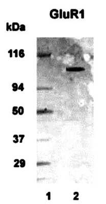
| Short Description | Rabbit polyclonal anti-GluR1 (Internal Region) for use in WB in Human, Mouse and Rat samples. Datasheet included with dilution recommendations, and related reagents. |
| Applications | WB |
| Host | Rabbit |
| Reactivity | Human/Mouse/Rat |
| Note | STRICTLY FOR FURTHER SCIENTIFIC RESEARCH USE ONLY (RUO). MUST NOT TO BE USED IN DIAGNOSTIC OR THERAPEUTIC APPLICATIONS. |
Product Properties
| Clonality | Polyclonal |
| Isotype | IgG |
| Conjugation | Unconjugated |
| Purification | Antigen Affinity Purified |
| Dilution Range | WB 1:500-1:1000 |
| Formulation | PBS + 0.02% NaN3 |
| Storage Instruction | Store at-20°C for up to 1 year from the date of receipt, and avoid repeat freeze-thaw cycles. |
Target Information
| Gene Symbol | GRIA1 |
| Gene ID | 2890 |
| Uniprot ID | GRIA1_HUMAN |
| Immunogen | Synthetic peptide corresponding to amino acids from an internal region of the human GluR1 alpha protein |
| Immunogen Region | Internal Region |
| Specificity | Reacts with internal residues of the human, rat and mouse GluR1 alpha protein. |
Additional Info
| Post Translational Modifications | Palmitoylated. Depalmitoylated by CPT1C and upon L-glutamate stimulation. ZDHHC3/GODZ specifically palmitoylates Cys-603, which leads to Golgi retention and decreased cell surface expression. In contrast, Cys-829 palmitoylation does not affect cell surface expression but regulates stimulation-dependent endocytosis. Phosphorylated at Ser-645. Phosphorylated at Ser-710 by PKC. Phosphorylated at Ser-849 by PKC, PKA and CAMK2. Phosphorylated at Ser-863 by PKC, PKA and PRKG2. Phosphorylation of Ser-863 is reduced by induction of long-term depression and increased by induction of long-term potentiation. |
| Function | Ionotropic glutamate receptor that functions as a ligand-gated cation channel, gated by L-glutamate and glutamatergic agonists such as alpha-amino-3-hydroxy-5-methyl-4-isoxazolepropionic acid (AMPA), quisqualic acid, and kainic acid. L-glutamate acts as an excitatory neurotransmitter at many synapses in the central nervous system. Binding of the excitatory neurotransmitter L-glutamate induces a conformation change, leading to the opening of the cation channel, and thereby converts the chemical signal to an electrical impulse upon entry of monovalent and divalent cations such as sodium and calcium. The receptor then desensitizes rapidly and enters in a transient inactive state, characterized by the presence of bound agonist. In the presence of CACNG2 or CACNG4 or CACNG7 or CACNG8, shows resensitization which is characterized by a delayed accumulation of current flux upon continued application of L-glutamate. Resensitization is blocked by CNIH2 through interaction with CACNG8 in the CACNG8-containing AMPA receptors complex. Calcium (Ca(2+)) permeability depends on subunits composition and, heteromeric channels containing edited GRIA2 subunit are calcium-impermeable. Also permeable to other divalents cations such as strontium(2+) and magnesium(2+) and monovalent cations such as potassium(1+) and lithium(1+). |
| Protein Name | Glutamate Receptor 1Glur-1Ampa-Selective Glutamate Receptor 1Glur-AGlur-K1Glutamate Receptor Ionotropic - Ampa 1 |
| Database Links | Reactome: R-HSA-204005Reactome: R-HSA-399710Reactome: R-HSA-399719Reactome: R-HSA-416993Reactome: R-HSA-438066Reactome: R-HSA-5694530Reactome: R-HSA-8849932Reactome: R-HSA-9620244 |
| Cellular Localisation | Cell MembraneMulti-Pass Membrane ProteinEndoplasmic Reticulum MembranePostsynaptic Cell MembranePostsynaptic Density MembraneCell ProjectionDendriteDendritic SpineEarly Endosome MembraneRecycling Endosome MembranePresynapseSynapseInteraction With Cacng2Cnih2 And Cnih3 Promotes Cell Surface ExpressionColocalizes With Pdlim4 In Early EndosomesDisplays A Somatodendritic Localization And Is Excluded From Axons In NeuronsLocalized To Cone Photoreceptor Pedicles |
| Alternative Antibody Names | Anti-Glutamate Receptor 1 antibodyAnti-Glur-1 antibodyAnti-Ampa-Selective Glutamate Receptor 1 antibodyAnti-Glur-A antibodyAnti-Glur-K1 antibodyAnti-Glutamate Receptor Ionotropic - Ampa 1 antibodyAnti-GRIA1 antibodyAnti-GLUA1 antibodyAnti-GLUH1 antibodyAnti-GLUR1 antibody |
Information sourced from Uniprot.org