


Anti-Collagen 1, alpha 1 telopeptide antibody (STJA0003600)
SPECIFICATIONS
ClonalityPolyclonal
HostRabbit
ConjugationUnconjugated
IsotypeIgG
ImmunogenSynthetic peptide corresponding to amino acid residues specific to the collagen 1, alpha 1 telopeptide conjugated to KLH.
General Information
| Short Description | Rabbit polyclonal anti-Collagen 1, alpha 1 telopeptide for use in WB and IHC in Human, Mouse, Rat, Sheep, Bovine, Canine, Chicken, Feline, Finch, Goat, Guinea Pig, Hamster, Horse, Non-Human Primates, Rabbit, Vole and Xenopus samples. Datasheet includ |
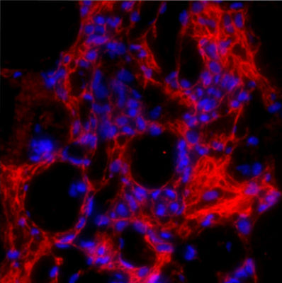
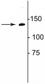
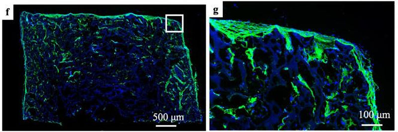
| Applications | WB/IHC |
| Host | Rabbit |
| Reactivity | Human/Mouse/Rat/Sheep/Bovine/Canine/Chicken/Feline/Finch/Goat/Guinea Pig/Hamster/Horse/Non-Human Primates/Rabbit/Vole/Xenopus |
| Note | STRICTLY FOR FURTHER SCIENTIFIC RESEARCH USE ONLY (RUO). MUST NOT TO BE USED IN DIAGNOSTIC OR THERAPEUTIC APPLICATIONS. |
Product Properties
| Clonality | Polyclonal |
| Isotype | IgG |
| Conjugation | Unconjugated |
| Purification | This antibody was antigen affinity purified from serum. |
| Dilution Range | WB 1:1000IHC 1:100-1:200 |
| Formulation | 100 ul in PBS |
| Storage Instruction | Store at-20°C for up to 1 year from the date of receipt, and avoid repeat freeze-thaw cycles. |
Target Information
| Gene Symbol | COL1A1 |
| Gene ID | 1277 |
| Uniprot ID | CO1A1_HUMAN |
| Immunogen | Synthetic peptide corresponding to amino acid residues specific to the collagen 1, alpha 1 telopeptide conjugated to KLH. |
Additional Info
| Tissue Specificity | Forms the fibrils of tendon, ligaments and bones. In bones the fibrils are mineralized with calcium hydroxyapatite. |
| Post Translational Modifications | Contains mostly 4-hydroxyproline. Proline residues at the third position of the tripeptide repeating unit (G-X-Y) are hydroxylated in some or all of the chains. Contains 3-hydroxyproline at a few sites. This modification occurs on the first proline residue in the sequence motif Gly-Pro-Hyp, where Hyp is 4-hydroxyproline. Lysine residues at the third position of the tripeptide repeating unit (G-X-Y) are 5-hydroxylated in some or all of the chains. O-glycosylated on hydroxylated lysine residues. The O-linked glycan consists of a Glc-Gal disaccharide. |
| Function | Type I collagen is a member of group I collagen (fibrillar forming collagen). |
| Protein Name | Collagen Alpha-1(I ChainAlpha-1 Type I Collagen |
| Database Links | Reactome: R-HSA-114604Reactome: R-HSA-1442490Reactome: R-HSA-1474244Reactome: R-HSA-1650814Reactome: R-HSA-198933Reactome: R-HSA-2022090Reactome: R-HSA-202733Reactome: R-HSA-216083Reactome: R-HSA-2214320Reactome: R-HSA-2243919Reactome: R-HSA-3000170Reactome: R-HSA-3000171Reactome: R-HSA-3000178Reactome: R-HSA-3000480Reactome: R-HSA-430116Reactome: R-HSA-75892Reactome: R-HSA-76009Reactome: R-HSA-8874081Reactome: R-HSA-8940973Reactome: R-HSA-8948216Reactome: R-HSA-9845619Reactome: R-HSA-9845620Reactome: R-HSA-9845621Reactome: R-HSA-9845622Reactome: R-HSA-9846298 |
| Cellular Localisation | SecretedExtracellular SpaceExtracellular Matrix |
| Alternative Antibody Names | Anti-Collagen Alpha-1(I Chain antibodyAnti-Alpha-1 Type I Collagen antibodyAnti-COL1A1 antibody |
Information sourced from Uniprot.org