| Host: |
Rabbit |
| Applications: |
WB/IHC/IF/IP |
| Reactivity: |
Human/Mouse/Rat |
| Note: |
STRICTLY FOR FURTHER SCIENTIFIC RESEARCH USE ONLY (RUO). MUST NOT TO BE USED IN DIAGNOSTIC OR THERAPEUTIC APPLICATIONS. |
| Short Description: |
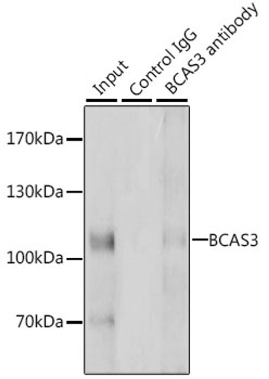
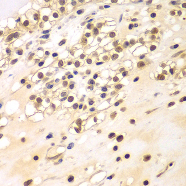
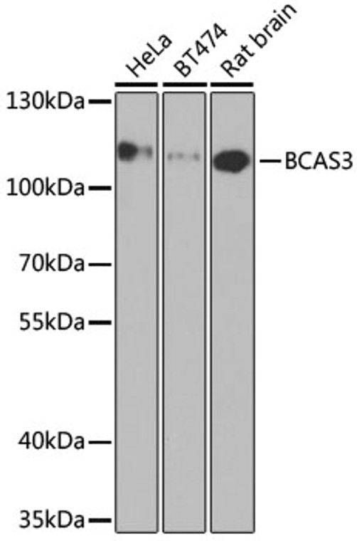
Rabbit polyclonal antibody anti-BCAS3 (674-913) is suitable for use in Western Blot, Immunohistochemistry, Immunofluorescence and Immunoprecipitation research applications. |
| Clonality: |
Polyclonal |
| Conjugation: |
Unconjugated |
| Isotype: |
IgG |
| Formulation: |
PBS with 0.02% Sodium Azide, 50% Glycerol, pH7.3. |
| Purification: |
Affinity purification |
| Dilution Range: |
WB 1:500-1:2000IHC-P 1:50-1:200IF/ICC 1:50-1:200IP 1:50-1:100 |
| Storage Instruction: |
Store at-20°C for up to 1 year from the date of receipt, and avoid repeat freeze-thaw cycles. |
| Gene Symbol: |
BCAS3 |
| Gene ID: |
54828 |
| Uniprot ID: |
BCAS3_HUMAN |
| Immunogen Region: |
674-913 |
| Immunogen: |
Recombinant fusion protein containing a sequence corresponding to amino acids 674-913 of human BCAS3 (NP_060149.3). |
| Immunogen Sequence: |
LAGLVPPGSPGPITRHGSYD SLASDHSGQEDEEWLSQVEI VTHTGPHRRLWMGPQFQFKT IHPSGQTTVISSSSSVLQSH GPSDTPQPLLDFDTDDLDLN SLRIQPVRSDPVSMPGSSRP VSDRRGVSTVIDAASGTFDR SVTLLEVCGSWPEGFGLRHM SSMEHTEEGLRERLADAMAE SPSRDVVGSGTELQREGSIE TLSNSSGSTSGSIPRNFDGY RSPLPTNESQPLSLFPTGF |
| Tissue Specificity | Expressed in stomach, liver, lung, kidney, prostate, testis, thyroid gland, adrenal gland, brain, heart, skeletal muscle, colon, spleen, small intestine, placenta, blood leukocyte and mammary epithelial cells. Expressed in undifferentiated ES cells. Expressed in blood islands and nascent blood vessels derived from differentiated ES cells into embryoid bodies (BD). Expressed in endothelial cells. Not detected in brain. Expressed in brain tumors (at protein level). Expressed in brain. Highly expressed in breast cancers and in glioma cell lines. |
| Function | Plays a role in angiogenesis. Participates in the regulation of cell polarity and directional endothelial cell migration by mediating both the activation and recruitment of CDC42 and the reorganization of the actin cytoskeleton at the cell leading edge. Promotes filipodia formation. Functions synergistically with PELP1 as a transcriptional coactivator of estrogen receptor-responsive genes. Stimulates histone acetyltransferase activity. Binds to chromatin. Plays a regulatory role in autophagic activity. In complex with PHAF1, associates with the preautophagosomal structure during both non-selective and selective autophagy. Probably binds phosphatidylinositol 3-phosphate (PtdIns3P) which would mediate the recruitment preautophagosomal structures. |
| Protein Name | Bcas3 Microtubule Associated Cell Migration FactorBreast Carcinoma-Amplified Sequence 3Gaob1 |
| Cellular Localisation | NucleusCytoplasmCytoskeletonPreautophagosomal StructureLocalizes In The Cytoplasm In Stationary CellsTranslocates From The Cytoplasm To The Leading Edge In Motile CellsColocalizes With Microtubules And Intermediate Filaments In Both Stationary And Motile CellsAssociates With ChromatinRecruited To Estrogen Receptor-Induced Promoters In A Pelp1-Dependent MannerThe Bcas3:Phaf1 Complex Is Recruited To The Preautophagosomal Structures Adjacent To The Damaged Mitochondria Upon Mitophagy In A Prkn-Pink1 Dependent Manner |
| Alternative Antibody Names | Anti-Bcas3 Microtubule Associated Cell Migration Factor antibodyAnti-Breast Carcinoma-Amplified Sequence 3 antibodyAnti-Gaob1 antibodyAnti-BCAS3 antibody |
Information sourced from Uniprot.org
12 months for antibodies. 6 months for ELISA Kits. Please see website T&Cs for further guidance


