


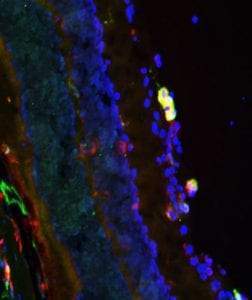
Anti-ARG1-ARG1 antibody (STJA0003835)
SPECIFICATIONS
ClonalityPolyclonal
HostChicken
ConjugationUnconjugated
IsotypeIgY
ImmunogenFull-length recombinant rat arginase I
General Information
| Short Description | Chicken polyclonal anti-Arginase I-ARG1 for use in WB and IHC in Guinea Pig, Human, Mouse and Rat samples. Datasheet included with dilution recommendations, and related reagents. |
| Applications | WB/IHC |
| Host | Chicken |
| Reactivity | Guinea Pig/Human/Mouse/Rat |
| Note | STRICTLY FOR FURTHER SCIENTIFIC RESEARCH USE ONLY (RUO). MUST NOT TO BE USED IN DIAGNOSTIC OR THERAPEUTIC APPLICATIONS. |
Product Properties
| Clonality | Polyclonal |
| Isotype | IgY |
| Conjugation | Unconjugated |
| Purification | This antibody was total igy fraction. |
| Dilution Range | WB 1:1000IHC 1:500 |
| Formulation | 100 µl in 10 mM HEPES (pH 7.5) , 150 mM NaCl, 100 µg per ml BSA and 50% Glycerol. |
| Storage Instruction | Store at-20°C for up to 1 year from the date of receipt, and avoid repeat freeze-thaw cycles. |
Target Information
| Gene Symbol | Arg1 |
| Gene ID | 29221 |
| Uniprot ID | ARGI1_RAT |
| Immunogen | Full-length recombinant rat arginase I |
Additional Info
| Tissue Specificity | Detected in liver (at protein level). |
| Function | Key element of the urea cycle converting L-arginine to urea and L-ornithine, which is further metabolized into metabolites proline and polyamides that drive collagen synthesis and bioenergetic pathways critical for cell proliferation, respectively.the urea cycle takes place primarily in the liver and, to a lesser extent, in the kidneys. Functions in L-arginine homeostasis in nonhepatic tissues characterized by the competition between nitric oxide synthase (NOS) and arginase for the available intracellular substrate arginine. Arginine metabolism is a critical regulator of innate and adaptive immune responses. Involved in an antimicrobial effector pathway in polymorphonuclear granulocytes (PMN). Upon PMN cell death is liberated from the phagolysosome and depletes arginine in the microenvironment leading to suppressed T cell and natural killer (NK) cell proliferation and cytokine secretion. In group 2 innate lymphoid cells (ILC2s) promotes acute type 2 inflammation in the lung and is involved in optimal ILC2 proliferation but not survival. Plays a role in the immune response of alternatively activated or M2 macrophages in processes such as wound healing and tissue regeneration, immune defense against multicellular pathogens and parasites, and immune suppression and allergic inflammation.the regulatory outcome seems to be organ specific. In tumor-infiltrating dendritic cells (DCs) and myeloid-derived suppressor cells (MDSCs) plays a role in suppression of T cell-mediated antitumor immunity. |
| Protein Name | Arginase-1Liver-Type ArginaseType I Arginase |
| Database Links | Reactome: R-RNO-6798695Reactome: -RNO-70635 |
| Cellular Localisation | CytoplasmCytoplasmic Granule |
| Alternative Antibody Names | Anti-Arginase-1 antibodyAnti-Liver-Type Arginase antibodyAnti-Type I Arginase antibodyAnti-Arg1 antibody |
Information sourced from Uniprot.org