| Host: |
Goat |
| Applications: |
Pep-ELISA/WB/IF/FC |
| Reactivity: |
Human/Mouse/Rat/Pig/Cow |
| Note: |
STRICTLY FOR FURTHER SCIENTIFIC RESEARCH USE ONLY (RUO). MUST NOT TO BE USED IN DIAGNOSTIC OR THERAPEUTIC APPLICATIONS. |
| Short Description: |
Goat polyclonal antibody anti-58KGolgi protein (Internal) /FTCD (Internal) is suitable for use in ELISA, Western Blot, Immunofluorescence and Flow Cytometry research applications. |
| Clonality: |
Polyclonal |
| Conjugation: |
Unconjugated |
| Isotype: |
IgG |
| Formulation: |
0.5 mg/ml in Tris saline, 0.02% sodium azide, pH7.3 with 0.5% bovine serum albumin. NA |
| Purification: |
Purified from goat serum by ammonium sulphate precipitation followed by antigen affinity chromatography using the immunizing peptide. |
| Concentration: |
0.5 mg/mL |
| Dilution Range: |
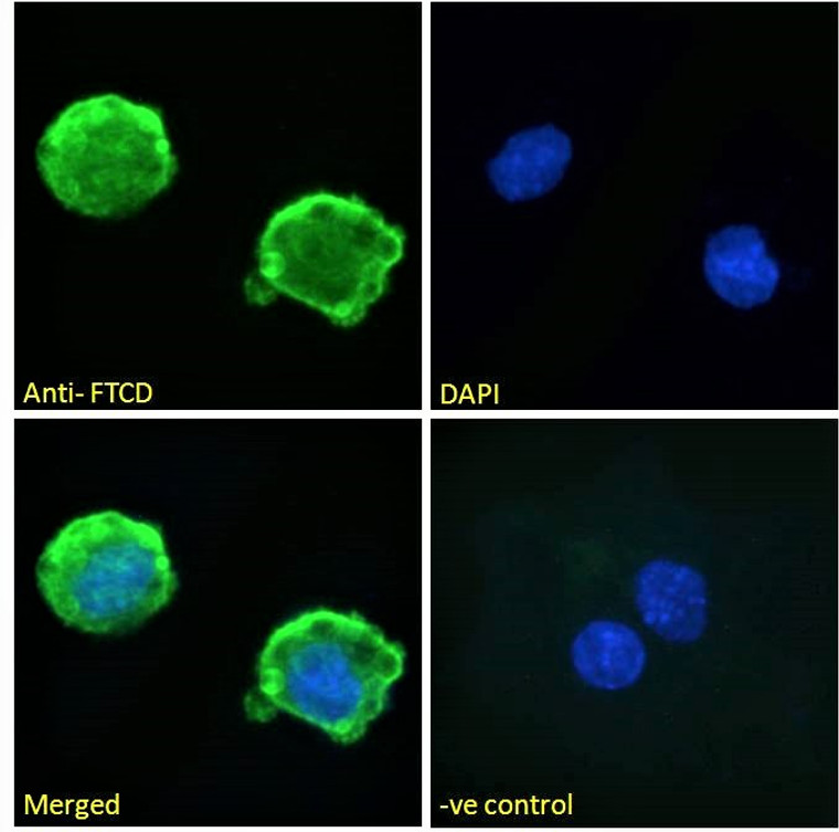
IF-Strong expression of the protein seen in the cytoplasm and plasma membranes of HeLa and in the membranes of HepG2 cells. 10µg/mlELISA-antibody detection limit dilution 1:64000. |
| Storage Instruction: |
Store at-20°C on receipt and minimise freeze-thaw cycles. |
| Gene Symbol: |
FTCD |
| Gene ID: |
10841 |
| Uniprot ID: |
FTCD_HUMAN |
| Immunogen Region: |
Internal |
| Accession Number: |
NP_006648.1; NP_996848.1; NP_001307341.1 |
| Specificity: |
Variants (NP_006648.1; NP_996848.1) encode the same protein. |
| Immunogen Sequence: |
CLREQGRGKDQPGRL |
| Protein Name | Formimidoyltransferase-CyclodeaminaseFormiminotransferase-CyclodeaminaseFtcdLchc1 Includes - Glutamate FormimidoyltransferaseGlutamate FormiminotransferaseGlutamate Formyltransferase - Formimidoyltetrahydrofolate CyclodeaminaseFormiminotetrahydrofolate Cyclodeaminase |
| Database Links | Reactome: R-HSA-70921 |
| Cellular Localisation | CytoplasmCytosolGolgi ApparatusCytoskeletonMicrotubule Organizing CenterCentrosomeCentrioleMore Abundantly Located Around The Mother Centriole |
| Alternative Antibody Names | Anti-Formimidoyltransferase-Cyclodeaminase antibodyAnti-Formiminotransferase-Cyclodeaminase antibodyAnti-Ftcd antibodyAnti-Lchc1 Includes - Glutamate Formimidoyltransferase antibodyAnti-Glutamate Formiminotransferase antibodyAnti-Glutamate Formyltransferase - Formimidoyltetrahydrofolate Cyclodeaminase antibodyAnti-Formiminotetrahydrofolate Cyclodeaminase antibodyAnti-FTCD antibody |
Information sourced from Uniprot.org
12 months for antibodies. 6 months for ELISA Kits. Please see website T&Cs for further guidance


