

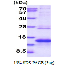

Human Melan-A/MART-1 protein (Recombinant) (His-Tag) (STJP018253)
SPECIFICATIONS
HostE.coli
ImmunogenHuman
General Information
| Short Description | Recombinant-Human Melan-A/MART-1-His-Tag protein was developed from e.coli and has a target region of His-Tag. For use in research applications. |
| Applications | SDS-PAGE |
| Host | E.coli |
| Note | STRICTLY FOR FURTHER SCIENTIFIC RESEARCH USE ONLY (RUO). MUST NOT TO BE USED IN DIAGNOSTIC OR THERAPEUTIC APPLICATIONS. |
Product Properties
| Concentration | 0.5 mg/mL |
| Formulation | Liquid in 20mM Tris-HCl buffer (pH 8.0) containing 0.15M NaCl, 20% Glycerol, 1mM DTT |
| Storage Instruction | For short term storage, keep at +2C to +8C for up to 1 week. For long term storage, aliquot and store at-20C, and avoid repeat freeze-thaw cycles. |
Target Information
| Gene Symbol | MLANA |
| Gene ID | 2315 |
| Uniprot ID | MAR1_HUMAN |
| Accession Number | NP_005502 |
| Immunogen | Human |
| Immunogen Region | 48-118aa |
| Immunogen Sequence | MGSSHHHHHH SSGLVPRGSH MGSCRRRNGY RALMDKSLHV GTQCALTRRC PQEGFDHRDS KVSLQEKNCE PVVPNAPPAY EKLSAEQSPP PYSP |
Additional Info
| Tissue Specificity | Expression is restricted to melanoma and melanocyte cell lines and retina. |
| Post Translational Modifications | Acylated. |
| Function | Involved in melanosome biogenesis by ensuring the stability of GPR143. Plays a vital role in the expression, stability, trafficking, and processing of melanocyte protein PMEL, which is critical to the formation of stage II melanosomes. |
| Protein Name | Melanoma Antigen Recognized By T-Cells 1Mart-1Antigen Lb39-AaAntigen Sk29-AaProtein Melan-A |
| Database Links | Reactome: R-HSA-9824585 |
| Cellular Localisation | Endoplasmic Reticulum MembraneSingle-Pass Type Iii Membrane ProteinGolgi ApparatusTrans-Golgi Network MembraneMelanosomeAlso Found In Small Vesicles And Tubules Dispersed Over The Entire CytoplasmA Small Fraction Of The Protein Is Inserted Into The Membrane In An Inverted OrientationInversion Of Membrane Topology Results In The Relocalization Of The Protein From A Predominant Golgi/Post-Golgi Area To The Endoplasmic ReticulumMelanoma Cells Expressing The Protein With An Inverted Membrane Topology Are More Effectively Recognized By Specific Cytolytic T-Lymphocytes Than Those Expressing The Protein In Its Native Membrane Orientation |
| Alternative Protein Names | Melanoma Antigen Recognized By T-Cells 1 proteinMart-1 proteinAntigen Lb39-Aa proteinAntigen Sk29-Aa proteinProtein Melan-A proteinMLANA proteinMART1 protein |
Information sourced from Uniprot.org