


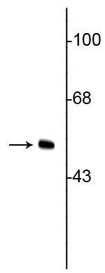
Anti-Vimentin antibody [2D1] (STJA0003824)
SPECIFICATIONS
ClonalityMonoclonal
HostMouse
ConjugationUnconjugated
IsotypeIgG2a
ImmunogenRecombinant human vimentin purification from E. coli.
General Information
| Short Description | Mouse monoclonal anti-Vimentin for use in WB, IHC and ICC in Human, Mouse, Rat, Bovine, Canine, Chicken, D.melanogaster, Feline, Finch, Fish, Goat, Guinea Pig, Hamster, Horse, Lizard, Non-Human Primates, Rabbit, Sheep, Vole, Xenopus and Zebrafish sam |
| Applications | WB/IHC/ICC |
| Host | Mouse |
| Reactivity | Human/Mouse/Rat/Bovine/Canine/Chicken/D.melanogaster/Feline/Finch/Fish/Goat/Guinea Pig/Hamster/Horse/Lizard/Non-Human Primates/Rabbit/Sheep/Vole/Xenopus/Zebrafish |
| Note | STRICTLY FOR FURTHER SCIENTIFIC RESEARCH USE ONLY (RUO). MUST NOT TO BE USED IN DIAGNOSTIC OR THERAPEUTIC APPLICATIONS. |
Product Properties
| Clonality | Monoclonal |
| Clone ID | 2D1 |
| Isotype | IgG2a |
| Conjugation | Unconjugated |
| Purification | This antibody was protein g purified culture from supernatant. |
| Dilution Range | WB 1:1000IHC 1:500-1:1000ICC 1:500-1:2000 |
| Formulation | 100 ul in PBS + 10 mM Sodium Azide. |
| Storage Instruction | Store at-20°C for up to 1 year from the date of receipt, and avoid repeat freeze-thaw cycles. |
Target Information
| Gene Symbol | VIM |
| Gene ID | 7431 |
| Uniprot ID | VIME_HUMAN |
| Immunogen | Recombinant human vimentin purification from E. coli. |
Additional Info
| Tissue Specificity | Highly expressed in fibroblasts, some expression in T- and B-lymphocytes, and little or no expression in Burkitt's lymphoma cell lines. Expressed in many hormone-independent mammary carcinoma cell lines. |
| Post Translational Modifications | Filament disassembly during mitosis is promoted by phosphorylation at Ser-55 as well as by nestin. One of the most prominent phosphoproteins in various cells of mesenchymal origin. Phosphorylation is enhanced during cell division, at which time vimentin filaments are significantly reorganized. Phosphorylation by PKN1 inhibits the formation of filaments. Phosphorylated at Ser-56 by CDK5 during neutrophil secretion in the cytoplasm. Phosphorylated by STK33. Phosphorylated on tyrosine residues by SRMS. O-glycosylated during cytokinesis at sites identical or close to phosphorylation sites, this interferes with the phosphorylation status. S-nitrosylation is induced by interferon-gamma and oxidatively-modified low-densitity lipoprotein (LDL(ox)) possibly implicating the iNOS-S100A8/9 transnitrosylase complex. |
| Function | Vimentins are class-III intermediate filaments found in various non-epithelial cells, especially mesenchymal cells. Vimentin is attached to the nucleus, endoplasmic reticulum, and mitochondria, either laterally or terminally. Plays a role in cell directional movement, orientation, cell sheet organization and Golgi complex polarization at the cell migration front. Protects SCRIB from proteasomal degradation and facilitates its localization to intermediate filaments in a cell contact-mediated manner. Involved with LARP6 in the stabilization of type I collagen mRNAs for CO1A1 and CO1A2. |
| Protein Name | Vimentin |
| Database Links | Reactome: R-HSA-264870Reactome: R-HSA-390522Reactome: R-HSA-6785807Reactome: R-HSA-9013422Reactome: R-HSA-9613829Reactome: R-HSA-9615710Reactome: R-HSA-9646399 |
| Cellular Localisation | CytoplasmCytoskeletonNucleus MatrixCell Membrane |
| Alternative Antibody Names | Anti-Vimentin antibodyAnti-VIM antibody |
Information sourced from Uniprot.org