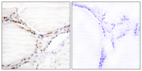
| Host: | Rabbit |
| Applications: | WB/IHC/IF/ELISA |
| Reactivity: | Human/Rat/Mouse |
| Note: | STRICTLY FOR FURTHER SCIENTIFIC RESEARCH USE ONLY (RUO). MUST NOT TO BE USED IN DIAGNOSTIC OR THERAPEUTIC APPLICATIONS. |
| Short Description : | Rabbit polyclonal antibody anti-Vitamin D3 receptor (181-230 aa) is suitable for use in Western Blot, Immunohistochemistry, Immunofluorescence and ELISA research applications. |
| Clonality : | Polyclonal |
| Conjugation: | Unconjugated |
| Isotype: | IgG |
| Formulation: | Liquid in PBS containing 50% Glycerol, 0.5% BSA and 0.02% Sodium Azide. |
| Purification: | The antibody was affinity-purified from rabbit antiserum by affinity-chromatography using epitope-specific immunogen. |
| Concentration: | 1 mg/mL |
| Dilution Range: | WB 1:500-1:2000IHC 1:100-1:300IF 1:200-1:1000ELISA 1:10000 |
| Storage Instruction: | Store at-20°C for up to 1 year from the date of receipt, and avoid repeat freeze-thaw cycles. |
| Gene Symbol: | VDR |
| Gene ID: | 7421 |
| Uniprot ID: | VDR_HUMAN |
| Immunogen Region: | 181-230 aa |
| Specificity: | VDR Polyclonal Antibody detects endogenous levels of VDR protein. |
| Immunogen: | The antiserum was produced against synthesized peptide derived from the human Vitamin D Receptor at the amino acid range 181-230 |
| Function | Nuclear receptor for calcitriol, the active form of vitamin D3 which mediates the action of this vitamin on cells. Enters the nucleus upon vitamin D3 binding where it forms heterodimers with the retinoid X receptor/RXR. The VDR-RXR heterodimers bind to specific response elements on DNA and activate the transcription of vitamin D3-responsive target genes. Plays a central role in calcium homeostasis. Also functions as a receptor for the secondary bile acid lithocholic acid (LCA) and its metabolites. |
| Protein Name | Vitamin D3 ReceptorVdr1 -25-Dihydroxyvitamin D3 ReceptorNuclear Receptor Subfamily 1 Group I Member 1 |
| Database Links | Reactome: R-HSA-196791Reactome: R-HSA-383280Reactome: R-HSA-4090294 |
| Cellular Localisation | NucleusCytoplasmLocalizes Mainly To The NucleusTranslocated Into The Nucleus Via Both Ligand-Dependent And Ligand-Independent PathwaysLigand-Independent Nuclear Translocation Is Mediated By Ipo4 |
| Alternative Antibody Names | Anti-Vitamin D3 Receptor antibodyAnti-Vdr antibodyAnti-1 -25-Dihydroxyvitamin D3 Receptor antibodyAnti-Nuclear Receptor Subfamily 1 Group I Member 1 antibodyAnti-VDR antibodyAnti-NR1I1 antibody |
Information sourced from Uniprot.org


