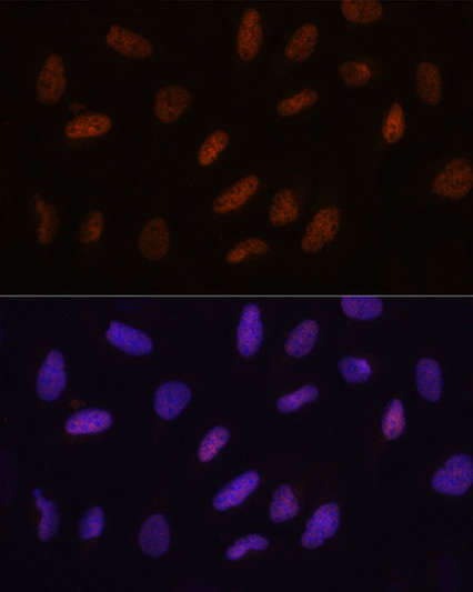
| Tissue Specificity | Expressed in colon, muscle, adrenal gland and peripheral blood lymphocytes. |
| Post Translational Modifications | Monoubiquitinated at Lys-994 by the DCX (DDB1-CUL4-X-box) E3 ubiquitin-protein ligase complex called CRL4(VprBP) or CUL4A-RBX1-DDB1-DCAF1/VPRBP complex.this modification promotes binding to DNA. |
| Function | Dioxygenase that catalyzes the conversion of the modified genomic base 5-methylcytosine (5mC) into 5-hydroxymethylcytosine (5hmC) and plays a key role in epigenetic chromatin reprogramming in the zygote following fertilization. Also mediates subsequent conversion of 5hmC into 5-formylcytosine (5fC), and conversion of 5fC to 5-carboxylcytosine (5caC). Conversion of 5mC into 5hmC, 5fC and 5caC probably constitutes the first step in cytosine demethylation. Selectively binds to the promoter region of target genes and contributes to regulate the expression of numerous developmental genes. In zygotes, DNA demethylation occurs selectively in the paternal pronucleus before the first cell division, while the adjacent maternal pronucleus and certain paternally-imprinted loci are protected from this process. Participates in DNA demethylation in the paternal pronucleus by mediating conversion of 5mC into 5hmC, 5fC and 5caC. Does not mediate DNA demethylation of maternal pronucleus because of the presence of DPPA3/PGC7 on maternal chromatin that prevents TET3-binding to chromatin. In addition to its role in DNA demethylation, also involved in the recruitment of the O-GlcNAc transferase OGT to CpG-rich transcription start sites of active genes, thereby promoting histone H2B GlcNAcylation by OGT. Binds preferentially to DNA containing cytidine-phosphate-guanosine (CpG) dinucleotides over CpH (H=A, T, and C), hemimethylated-CpG and hemimethylated-hydroxymethyl-CpG. |
| Protein Name | Methylcytosine Dioxygenase Tet3 |
| Database Links | Reactome: R-HSA-5221030Reactome: R-HSA-9821002 |
| Cellular Localisation | NucleusCytoplasmChromosomeAt The Zygotic StageLocalizes In The Male PronucleusWhile It Localizes To The Cytoplasm At Other Preimplantation StagesBinds To The Promoter Of Target GenesClose To The Transcription Start Site |
| Alternative Antibody Names | Anti-Methylcytosine Dioxygenase Tet3 antibodyAnti-TET3 antibodyAnti-CXXC10 antibodyAnti-KIAA0401 antibody |
Information sourced from Uniprot.org


