

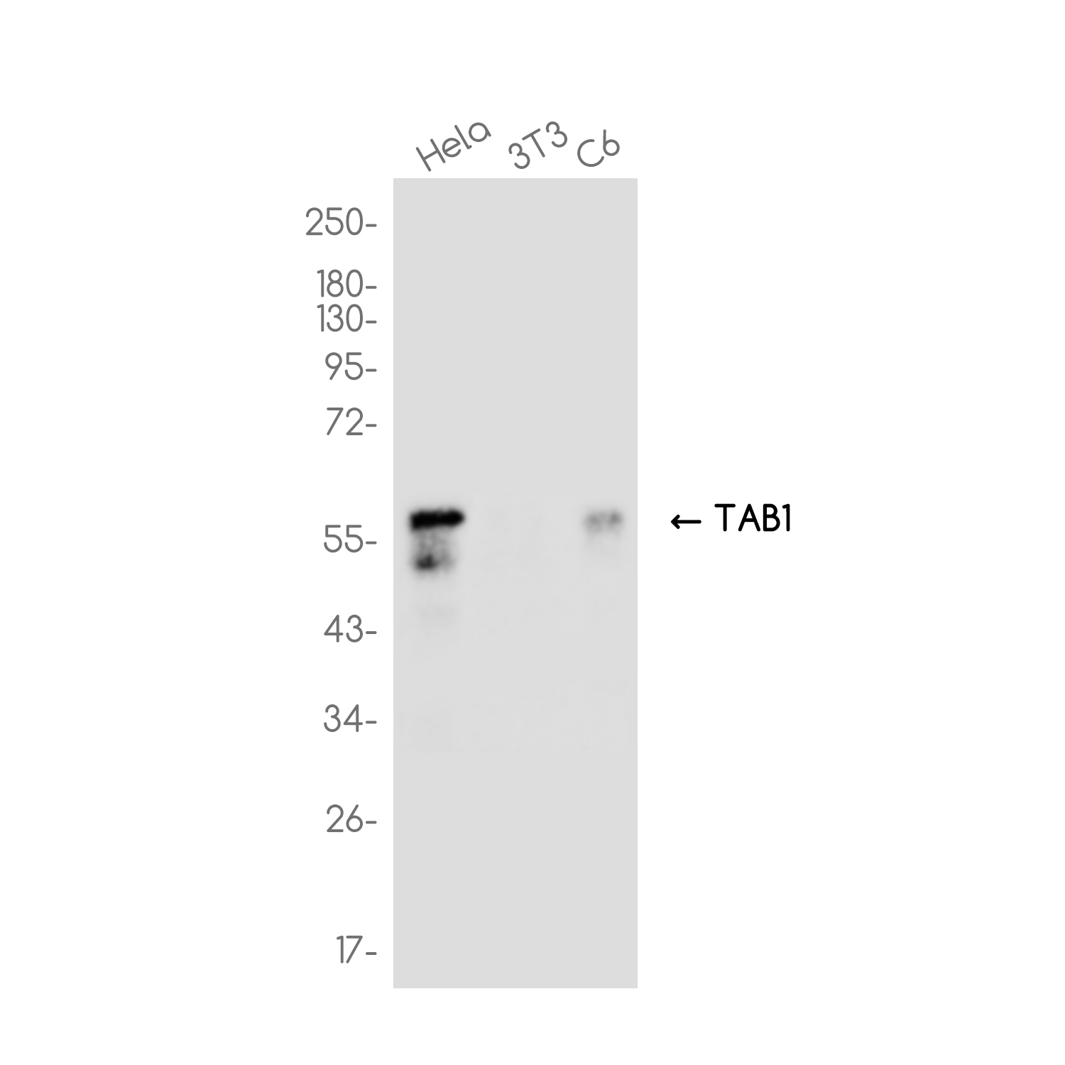
Anti-TAB1 antibody [R03-7J9] (STJA0036965)
SPECIFICATIONS
ClonalityMonoclonal
HostRabbit
ConjugationUnconjugated
IsotypeIgG
ImmunogenA synthesized peptide derived from human TAB1
General Information
| Short Description | Rabbit monoclonal anti-TAB1 for use in WB, IHC-P, ICC, IF, IP and FC in Human, Mouse and Rat samples. Datasheet included with dilution recommendations, and related reagents. |
| Applications | WB/IHC-P/ICC/IF/IP/FC |
| Host | Rabbit |
| Reactivity | Human/Mouse/Rat |
| Note | STRICTLY FOR FURTHER SCIENTIFIC RESEARCH USE ONLY (RUO). MUST NOT TO BE USED IN DIAGNOSTIC OR THERAPEUTIC APPLICATIONS. |
Product Properties
| Clonality | Monoclonal |
| Clone ID | R03-7J9 |
| Isotype | IgG |
| Conjugation | Unconjugated |
| Purification | Affinity Chromatography |
| Dilution Range | WB 1:500-1:1000IHC 1:50-1:100IF 1:50-1:200IP 1:40FC 1:50-1:100 |
| Formulation | Rabbit IgG in phosphate buffered saline, pH 7.4, 150mM NaCl, 0.02% sodium azide and 50% glycerol. |
| Storage Instruction | Store at 4°C short term. Aliquot and store at-20°C long term. Avoid freeze/thaw cycles. |
Target Information
| Gene Symbol | TAB1 |
| Gene ID | 10454 |
| Uniprot ID | TAB1_HUMAN |
| Immunogen | A synthesized peptide derived from human TAB1 |
Additional Info
| Tissue Specificity | Ubiquitous. |
| Post Translational Modifications | Phosphorylated at all three sites Ser-423, Thr-431 and Ser-438 by MAPK14 when cells were exposed to cellular stresses, or stimulated with TNF-alpha, IL1 or LPS. These phosphorylations inhibit TAK1 activation by a feedback control mechanism. Dephosphorylated by DUSP14 at Ser-438, leading to TAB1-MAP3K7/TAK1 complex inactivation in T-cells. Ubiquitinated by MAP3K1 with 'Lys-63'-linked polyubiquitin.leading to activation of TAK1 and of JNK and p38 MAP kinases following EGF and TGF-beta stimulation. Ubiquitinated by ITCH with 'Lys-48'-linked polyubiquitin.leading to proteasomal degradation. Ubiquitinated by RNF114 during maternal-to-zygotic transition.leading to degradation. (Microbial infection) Deubiquitinated by Y.enterocolitica YopP. O-GlcNAcylated at Ser-395 by OGT is required for full MAP3K7/TAK1 activation upon stimulation with IL-1 or osmotic stress. Deglycosylated at Ser-395 by OGA. |
| Function | Key adapter protein that plays an essential role in JNK and NF-kappa-B activation and proinflammatory cytokines production in response to stimulation with TLRs and cytokines. Mechanistically, associates with the catalytic domain of MAP3K7/TAK1 to trigger MAP3K7/TAK1 autophosphorylation leading to its full activation. Similarly, associates with MAPK14 and triggers its autophosphorylation and subsequent activation. In turn, MAPK14 phosphorylates TAB1 and inhibits MAP3K7/TAK1 activation in a feedback control mechanism. Also plays a role in recruiting MAPK14 to the TAK1 complex for the phosphorylation of the TAB2 and TAB3 regulatory subunits. |
| Protein Name | Tgf-Beta-Activated Kinase 1 And Map3k7-Binding Protein 1Mitogen-Activated Protein Kinase Kinase Kinase 7-Interacting Protein 1Tgf-Beta-Activated Kinase 1-Binding Protein 1Tak1-Binding Protein 1 |
| Database Links | Reactome: R-HSA-168638Reactome: R-HSA-2871837Reactome: R-HSA-445989Reactome: R-HSA-450302Reactome: R-HSA-450321Reactome: R-HSA-5357956Reactome: R-HSA-5607764Reactome: R-HSA-5689880Reactome: R-HSA-9014325Reactome: R-HSA-9020702Reactome: R-HSA-937042Reactome: R-HSA-937072Reactome: R-HSA-9645460Reactome: R-HSA-9705671Reactome: R-HSA-975163 |
| Cellular Localisation | CytoplasmCytosolEndoplasmic Reticulum MembranePeripheral Membrane ProteinCytoplasmic SideRecruited To The Endoplasmic Reticulum Following Interaction With Sting1 |
| Alternative Antibody Names | Anti-Tgf-Beta-Activated Kinase 1 And Map3k7-Binding Protein 1 antibodyAnti-Mitogen-Activated Protein Kinase Kinase Kinase 7-Interacting Protein 1 antibodyAnti-Tgf-Beta-Activated Kinase 1-Binding Protein 1 antibodyAnti-Tak1-Binding Protein 1 antibodyAnti-TAB1 antibodyAnti-MAP3K7IP1 antibody |
Information sourced from Uniprot.org