| Host: | Rabbit |
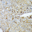
| Applications: | WB/IHC-P/ELISA |
| Reactivity: | Human/Mouse |
| Note: | STRICTLY FOR FURTHER SCIENTIFIC RESEARCH USE ONLY (RUO). MUST NOT TO BE USED IN DIAGNOSTIC OR THERAPEUTIC APPLICATIONS. |
| Clonality : | Polyclonal |
| Conjugation: | Unconjugated |
| Isotype: | IgG |
| Formulation: | PBS with 0.05% Proclin300, 50% Glycerol, pH 7.3. |
| Purification: | Affinity purification |
| Concentration: | Lot specific |
| Dilution Range: | WB:1:500-1:2000IHC-P:1:50-1:200ELISA:Recommended starting concentration is 1 Mu g/mL. Please optimize the concentration based on your specific assay requirements. |
| Storage Instruction: | Store at-20°C for up to 1 year from the date of receipt, and avoid repeat freeze-thaw cycles. |
| Gene Symbol: | SDC1 |
| Gene ID: | 6382 |
| Uniprot ID: | SDC1_HUMAN |
| Immunogen Region: | 25-240 |
| Specificity: | Recombinant fusion protein containing a sequence corresponding to amino acids 25-240 of human CD138/Syndecan-1 (NP_002988.4). |
| Immunogen Sequence: | VATNLPPEDQDGSGDDSDNF SGSGAGALQDITLSQQTPST WKDTQLLTAIPTSPEPTGLE ATAASTSTLPAGEGPKEGEA VVLPEVEPGLTAREQEATPR PRETTQLPTTHLASTTTATT AQEPATSHPHRDMQPGHHET STPAGPSQADLHTPHTEDGG PSATERAAEDGASSQLPAAE GSGEQDFTFETSGENTAVVA VEPDRRNQSPVDQGAT |
| Tissue Specificity | Detected in placenta (at protein level). Detected in fibroblasts (at protein level). |
| Post Translational Modifications | Shedding is enhanced by a number of factors such as heparanase, thrombin or EGF. Also by stress and wound healing. PMA-mediated shedding is inhibited by TIMP3. |
| Function | Cell surface proteoglycan that contains both heparan sulfate and chondroitin sulfate and that links the cytoskeleton to the interstitial matrix. Regulates exosome biogenesis in concert with SDCBP and PDCD6IP. Able to induce its own expression in dental mesenchymal cells and also in the neighboring dental epithelial cells via an MSX1-mediated pathway. |
| Protein Name | Syndecan-1Synd1Cd Antigen Cd138 |
| Database Links | Reactome: R-HSA-1971475Reactome: R-HSA-2022928Reactome: R-HSA-2024096Reactome: R-HSA-202733Reactome: R-HSA-3000170Reactome: R-HSA-3560783Reactome: R-HSA-3560801Reactome: R-HSA-3656237Reactome: R-HSA-3656253Reactome: R-HSA-4420332Reactome: R-HSA-449836Reactome: R-HSA-9694614Reactome: R-HSA-975634Reactome: R-HSA-9820960Reactome: R-HSA-9833110 |
| Cellular Localisation | MembraneSingle-Pass Type I Membrane ProteinSecretedExtracellular ExosomeShedding Of The Ectodomain Produces A Soluble Form |
| Alternative Antibody Names | Anti-Syndecan-1 antibodyAnti-Synd1 antibodyAnti-Cd Antigen Cd138 antibodyAnti-SDC1 antibodyAnti-SDC antibody |
Information sourced from Uniprot.org


