


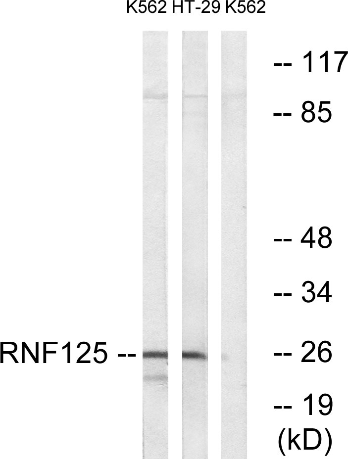
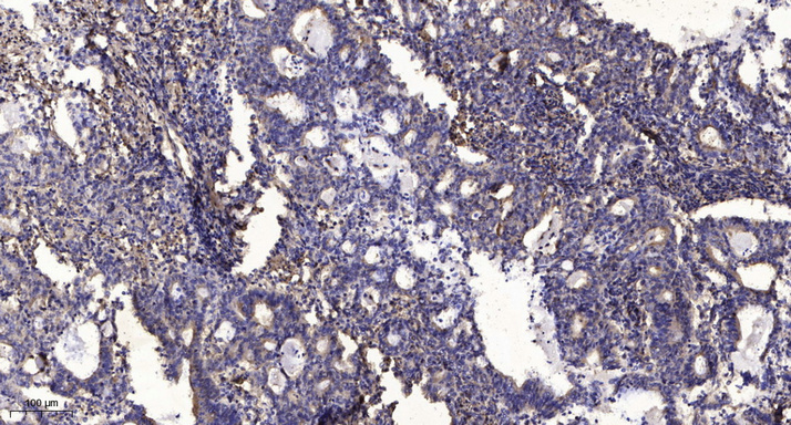
Anti-RNF125 antibody (131-180 aa) (STJ96080)
SPECIFICATIONS
ClonalityPolyclonal
HostRabbit
ConjugationUnconjugated
IsotypeIgG
ImmunogenThe antiserum was produced against synthesized peptide derived from the human RNF125 at the amino acid range 131-180
General Information
| Short Description | Rabbit polyclonal anti-E3 ubiquitin-protein ligase RNF125 (131-180 aa) for use in WB, IHC, IF and ELISA in Human, Mouse and Rat samples. Datasheet included with dilution recommendations, and related reagents. |
| Applications | WB/IHC/IF/ELISA |
| Host | Rabbit |
| Reactivity | Human/Mouse/Rat |
| Note | STRICTLY FOR FURTHER SCIENTIFIC RESEARCH USE ONLY (RUO). MUST NOT TO BE USED IN DIAGNOSTIC OR THERAPEUTIC APPLICATIONS. |
Product Properties
| Clonality | Polyclonal |
| Isotype | IgG |
| Conjugation | Unconjugated |
| Concentration | 1 mg/mL |
| Purification | The antibody was affinity-purified from rabbit antiserum by affinity-chromatography using epitope-specific immunogen. |
| Dilution Range | WB 1:500-1:2000IHC 1:100-1:300ELISA 1:10000IF 1:50-200 |
| Formulation | Liquid in PBS containing 50% Glycerol, 0.5% BSA and 0.02% Sodium Azide. |
| Storage Instruction | Store at-20°C for up to 1 year from the date of receipt, and avoid repeat freeze-thaw cycles. |
Target Information
| Gene Symbol | RNF125 |
| Gene ID | 54941 |
| Uniprot ID | RN125_HUMAN |
| Immunogen | The antiserum was produced against synthesized peptide derived from the human RNF125 at the amino acid range 131-180 |
| Immunogen Region | 131-180 aa |
| Specificity | TRAC-1 Polyclonal Antibody detects endogenous levels of TRAC-1 protein. |
Additional Info
| Post Translational Modifications | Autoubiquitinated, leading to its subsequent proteasomal degradation. |
| Function | E3 ubiquitin-protein ligase that mediates ubiquitination and subsequent proteasomal degradation of target proteins, such as RIGI, MAVS/IPS1, IFIH1/MDA5, JAK1 and p53/TP53. Acts as a negative regulator of type I interferon production by mediating ubiquitination of RIGI at 'Lys-181', leading to RIGI degradation. Mediates ubiquitination and subsequent degradation of p53/TP53. Mediates ubiquitination and subsequent degradation of JAK1. Acts as a positive regulator of T-cell activation. |
| Protein Name | E3 Ubiquitin-Protein Ligase Rnf125Ring Finger Protein 125T-Cell Ring Activation Protein 1Trac-1 |
| Database Links | Reactome: R-HSA-936440 |
| Cellular Localisation | Golgi Apparatus MembraneLipid-AnchorShows A Reticular Staining Pattern Within The Cell And Is Probably Expressed At Other Intracellular Membranes In Addition To The Golgi MembraneNot Detected At The Plasma Membrane |
| Alternative Antibody Names | Anti-E3 Ubiquitin-Protein Ligase Rnf125 antibodyAnti-Ring Finger Protein 125 antibodyAnti-T-Cell Ring Activation Protein 1 antibodyAnti-Trac-1 antibodyAnti-RNF125 antibody |
Information sourced from Uniprot.org