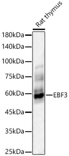
| Tissue Specificity | Expressed in kidney, heart, liver, and skeletal muscle. Also present in placenta and peripheral blood leukocytes. Not detected in colon, thymus, spleen and small intestine. In lung, expressed in bronchial epithelial cells and alveolar macrophages, but scarcely detected in alveolar epithelium, arterial walls and interstitial fibroblasts (at protein level). In joints of osteoarthritis and rheumatoid arthritis, expressed in endothelial cells (at protein level). In normal heart, detected in some vessels. In myocardial tissues with acute infarction, expressed in vascular endothelial cells adjacent to cardiomyocytes and those in lesions with granulation. Expression in cardiomyocytes is scarce (at protein level). In uterus, breast and colon cancers, detected in tumor cells and neighboring microvascular endothelium, but not in normal glandular tissues (at protein level). Expressed in dermal resting mast cells (at protein level) and pulmonary mast cells. Expressed in neuronal fibers (at protein level). Highly expressed in dorsal root ganglia neurons (at protein level). Expressed in Purkinje cells in cerebellum (at protein level). In stomach is preferentially expressed in neuronal fibers and in microvascular endothelium. Sparsely expressed in normal aorta (at protein level). Highly expressed in macrophages and smooth muscle cells in aorta with atheroma. |
| Post Translational Modifications | N-glycosylation does not affect the catalytic activity, but is required for proper secretion. A nonglycosylated form is observed in several cell types. In several cell types, the N- and C-termini are cleaved off. |
| Function | Secretory calcium-dependent phospholipase A2 that primarily targets extracellular phospholipids. Hydrolyzes the ester bond of the fatty acyl group attached at sn-2 position of phospholipids without apparent head group selectivity. Contributes to phospholipid remodeling of low-density lipoprotein (LDL) and high-density lipoprotein (HDL) particles. Hydrolyzes LDL phospholipids releasing unsaturated fatty acids that regulate macrophage differentiation toward foam cells. May act in an autocrine and paracrine manner. Secreted by immature mast cells, acts on nearby fibroblasts upstream to PTDGS to synthesize prostaglandin D2 (PGD2), which in turn promotes mast cell maturation and degranulation via PTGDR. Secreted by epididymal epithelium, acts on immature sperm cells within the duct, modulating the degree of unsaturation of the fatty acyl components of phosphatidylcholines required for acrosome assembly and sperm cell motility. Facilitates the replacement of fatty acyl chains in phosphatidylcholines in sperm membranes from omega-6 and omega-9 to omega-3 polyunsaturated fatty acids (PUFAs). Coupled to lipoxygenase pathway, may process omega-6 PUFAs to generate oxygenated lipid mediators in the male reproductive tract. At pericentrosomal preciliary compartment, negatively regulates ciliogenesis likely by regulating endocytotic recycling of ciliary membrane protein. Coupled to cyclooxygenase pathway provides arachidonate to generate prostaglandin E2 (PGE2), a potent immunomodulatory lipid in inflammation and tumorigenesis. At colonic epithelial barrier, preferentially hydrolyzes phospholipids having arachidonate and docosahexaenoate at sn-2 position, contributing to the generation of oxygenated metabolites involved in colonic stem cell homeostasis. Releases C16:0 and C18:0 lysophosphatidylcholine subclasses from neuron plasma membranes and promotes neurite outgrowth and neuron survival. |
| Protein Name | Group 3 Secretory Phospholipase A2Group Iii Secretory Phospholipase A2Giii Spla2Spla2-IiiPhosphatidylcholine 2-Acylhydrolase 3 |
| Database Links | Reactome: R-HSA-1482788Reactome: R-HSA-1482839Reactome: R-HSA-1482925 |
| Cellular Localisation | SecretedCell MembraneCytoplasmCytoskeletonMicrotubule Organizing CenterCentrosomeCentrioleRecycling EndosomeLocalized At Pericentrosomal Preciliary Compartment |
| Alternative Antibody Names | Anti-Group 3 Secretory Phospholipase A2 antibodyAnti-Group Iii Secretory Phospholipase A2 antibodyAnti-Giii Spla2 antibodyAnti-Spla2-Iii antibodyAnti-Phosphatidylcholine 2-Acylhydrolase 3 antibodyAnti-PLA2G3 antibody |
Information sourced from Uniprot.org


