
. 2, STJ194749")

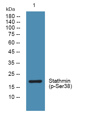
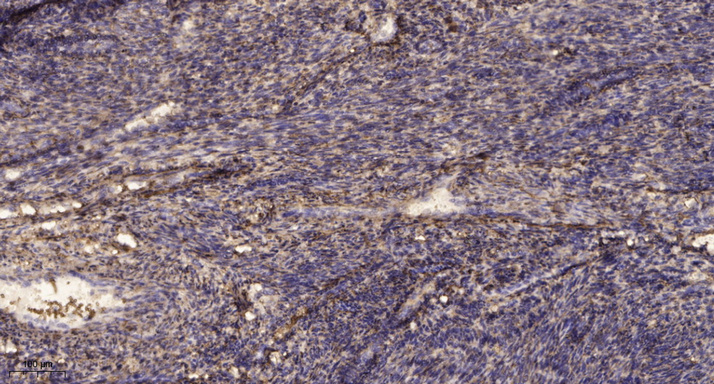
Anti-Phospho-STMN2-Ser38 antibody (STJ194749)
SPECIFICATIONS
ClonalityPolyclonal
HostRabbit
ConjugationUnconjugated
IsotypeIgG
ImmunogenSynthesized phosho peptide around human Stathmin (Ser38)
General Information
| Short Description | Rabbit polyclonal anti-Phospho-Stathmin-2-Ser38 for use in WB and IHC in Human, Rat and Mouse samples. Datasheet included with dilution recommendations, and related reagents. |
| Applications | WB/IHC |
| Host | Rabbit |
| Reactivity | Human/Rat/Mouse |
| Note | STRICTLY FOR FURTHER SCIENTIFIC RESEARCH USE ONLY (RUO). MUST NOT TO BE USED IN DIAGNOSTIC OR THERAPEUTIC APPLICATIONS. |
Product Properties
| Clonality | Polyclonal |
| Isotype | IgG |
| Conjugation | Unconjugated |
| Concentration | 1 mg/mL |
| Purification | The antibody was affinity-purified from rabbit serum by affinity-chromatography using specific immunogen. |
| Dilution Range | WB 1:500-2000IHC-P 1:50-300 |
| Formulation | Liquid in PBS containing 50% Glycerol, 0.5% BSA and 0.02% Sodium Azide. |
| Storage Instruction | Store at-20°C for up to 1 year from the date of receipt, and avoid repeat freeze-thaw cycles. |
Target Information
| Gene Symbol | STMN2 |
| Gene ID | 11075 |
| Uniprot ID | STMN2_HUMAN |
| Immunogen | Synthesized phosho peptide around human Stathmin (Ser38) |
| Specificity | This antibody detects endogenous levels of Human Stathmin (phospho-Ser38) |
Additional Info
| Post Translational Modifications | Sumoylated. Phosphorylated mostly by MAPK8, but also by MAPK9 and MAPK10 in the developing brain cortex. N-terminal palmitoylation promotes specific anchoring to the cytosolic leaflet of Golgi membranes and subsequent vesicular trafficking along dendrites and axons. Neuronal Stathmins are substrates for palmitoyltransferases ZDHHC3, ZDHHC7 and ZDHHC15. |
| Function | Regulator of microtubule stability. When phosphorylated by MAPK8, stabilizes microtubules and consequently controls neurite length in cortical neurons. In the developing brain, negatively regulates the rate of exit from multipolar stage and retards radial migration from the ventricular zone. |
| Protein Name | Stathmin-2Superior Cervical Ganglion-10 ProteinProtein Scg10 |
| Database Links | Reactome: R-HSA-9696273 |
| Cellular Localisation | CytoplasmPerinuclear RegionCell ProjectionGrowth ConeMembranePeripheral Membrane ProteinCytoplasmic SideAxonGolgi ApparatusEndosomeLamellipodiumAssociated With Punctate Structures In The Perinuclear CytoplasmAxonsAnd Growth Cones Of Developing NeuronsScg10 Exists In Both Soluble And Membrane-Bound FormsColocalized With Cib1 In Neurites Of Developing Hippocampal Primary NeuronsColocalized With Cib1 In The Cell BodyNeuritis And Growth Cones Of NeuronsColocalized With Cib1 To The Leading Edge Of Lamellipodia |
| Alternative Antibody Names | Anti-Stathmin-2 antibodyAnti-Superior Cervical Ganglion-10 Protein antibodyAnti-Protein Scg10 antibodyAnti-STMN2 antibodyAnti-SCG10 antibodyAnti-SCGN10 antibody |
Information sourced from Uniprot.org