
 antibody. STJ98682")

 for Immunogen Phosphopeptide (Phospho-left) and Non-Phosphopeptide STJ98682")

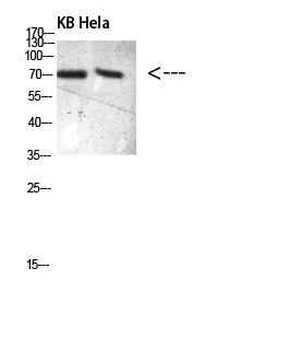
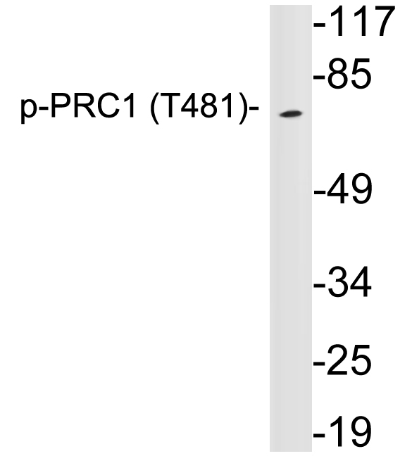
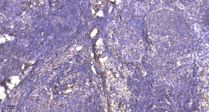
Anti-Phospho-PRC1-Thr481 antibody (481 aa) (STJ98682)
SPECIFICATIONS
ClonalityPolyclonal
HostRabbit
ConjugationUnconjugated
IsotypeIgG
ImmunogenSynthetic Phospho peptide from the human protein at the amino acid range 481
General Information
| Short Description | Rabbit polyclonal anti-Phospho-Protein regulator of cytokinesis 1-Thr481 (481 aa) for use in WB and IHC in Human, Rat and Mouse samples. Datasheet included with dilution recommendations, and related reagents. |
| Applications | WB/IHC |
| Host | Rabbit |
| Reactivity | Human/Rat/Mouse |
| Note | STRICTLY FOR FURTHER SCIENTIFIC RESEARCH USE ONLY (RUO). MUST NOT TO BE USED IN DIAGNOSTIC OR THERAPEUTIC APPLICATIONS. |
Product Properties
| Clonality | Polyclonal |
| Isotype | IgG |
| Conjugation | Unconjugated |
| Concentration | 1 mg/mL |
| Purification | The antibody was affinity-purified from rabbit antiserum by affinity-chromatography using epitope-specific immunogen. |
| Dilution Range | WB 1:500-2000IHC-P 1:50-300 |
| Formulation | Liquid in PBS containing 50% Glycerol, 0.5% BSA and 0.02% Sodium Azide. |
| Storage Instruction | Store at-20°C for up to 1 year from the date of receipt, and avoid repeat freeze-thaw cycles. |
Target Information
| Gene Symbol | PRC1 |
| Gene ID | 9055 |
| Uniprot ID | PRC1_HUMAN |
| Immunogen | Synthetic Phospho peptide from the human protein at the amino acid range 481 |
| Immunogen Region | 481 aa |
| Specificity | The antibody detects endogenous PRC1Phospho occurs at Thr481 |
Additional Info
| Post Translational Modifications | Phosphorylation by CDK1 in early mitosis holds PRC1 in an inactive monomeric state, during the metaphase to anaphase transition, PRC1 is dephosphorylated, promoting interaction with KIF4A, which then translocates PRC1 along mitotic spindles to the plus ends of antiparallel interdigitating microtubules. Dephosphorylation also promotes MT-bundling activity by allowing dimerization. Phosphorylation by CDK1 prevents PLK1-binding: upon degradation of CDK1 at anaphase and dephosphorylation, it is then phosphorylated by PLK1, leading to cytokinesis. |
| Function | Key regulator of cytokinesis that cross-links antiparrallel microtubules at an average distance of 35 nM. Essential for controlling the spatiotemporal formation of the midzone and successful cytokinesis. Required for KIF14 localization to the central spindle and midbody. Required to recruit PLK1 to the spindle. Stimulates PLK1 phosphorylation of RACGAP1 to allow recruitment of ECT2 to the central spindle. Acts as an oncogene for promoting bladder cancer cells proliferation, apoptosis inhibition and carcinogenic progression. |
| Protein Name | Protein Regulator Of Cytokinesis 1 |
| Database Links | Reactome: R-HSA-5625900 |
| Cellular Localisation | NucleusCytoplasmCytoskeletonSpindle PoleMidbodyChromosomeColocalized With Kif20b In The Nucleus Of Bladder Carcinoma Cells At The InterphaseColocalized With Kif20b In Bladder Carcinoma Cells At ProphaseMetaphaseEarly AnaphaseAt The Midzone In Late Anaphase And At The Contractile Ring In TelophasePredominantly Localized To The Nucleus Of Interphase CellsDuring Mitosis Becomes Associated With The Mitotic Spindle Poles And Localizes With The Cell Midbody During CytokinesisCo-Localizes With Prc1 In Early Mitosis And At The Spindle Midzone From Anaphase B To Telophase |
| Alternative Antibody Names | Anti-Protein Regulator Of Cytokinesis 1 antibodyAnti-PRC1 antibody |
Information sourced from Uniprot.org