 Antibody. STJ90689")
. STJ90689")
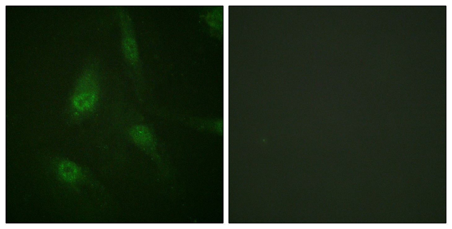
 Antibody. The picture on the right is blocked STJ90689")
 Antibody. STJ90689")
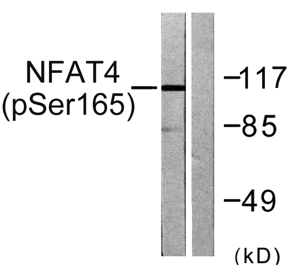
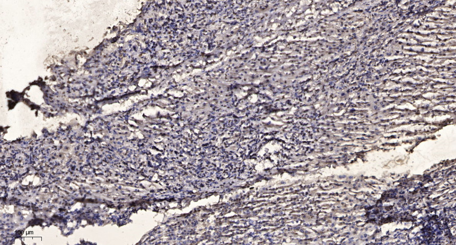
Anti-Phospho-NFATC3-Ser165 antibody (131-180 aa) (STJ90689)
SPECIFICATIONS
ClonalityPolyclonal
HostRabbit
ConjugationUnconjugated
IsotypeIgG
ImmunogenThe antiserum was produced against synthesized peptide derived from the human NFAT4 around the phosphorylation site of Ser165 at the amino acid range 131-180
General Information
| Short Description | Rabbit polyclonal anti-Phospho-Nuclear factor of activated T-cells, cytoplasmic 3-Ser165 (131-180 aa) for use in WB, IHC, IF and ELISA in Human and Mouse samples. Datasheet included with dilution recommendations, and related reagents. |
| Applications | WB/IHC/IF/ELISA |
| Host | Rabbit |
| Reactivity | Human/Mouse |
| Note | STRICTLY FOR FURTHER SCIENTIFIC RESEARCH USE ONLY (RUO). MUST NOT TO BE USED IN DIAGNOSTIC OR THERAPEUTIC APPLICATIONS. |
Product Properties
| Clonality | Polyclonal |
| Isotype | IgG |
| Conjugation | Unconjugated |
| Concentration | 1 mg/mL |
| Purification | The antibody was affinity-purified from rabbit antiserum by affinity-chromatography using epitope-specific immunogen. |
| Dilution Range | WB 1:500-1:2000IHC 1:100-1:300IF 1:200-1:1000ELISA 1:5000 |
| Formulation | Liquid in PBS containing 50% Glycerol, 0.5% BSA and 0.02% Sodium Azide. |
| Storage Instruction | Store at-20°C for up to 1 year from the date of receipt, and avoid repeat freeze-thaw cycles. |
Target Information
| Gene Symbol | NFATC3 |
| Gene ID | 4775 |
| Uniprot ID | NFAC3_HUMAN |
| Immunogen | The antiserum was produced against synthesized peptide derived from the human NFAT4 around the phosphorylation site of Ser165 at the amino acid range 131-180 |
| Immunogen Region | 131-180 aa |
| Specificity | Phospho-NFATc3 (S165) Polyclonal Antibody detects endogenous levels of NFATc3 protein only when phosphorylated at S165. |
Additional Info
| Post Translational Modifications | Ubiquitinated by STUB1/CHIP, leading to proteasomal degradation. Phosphorylated by NFATC-kinase.dephosphorylated by calcineurin. |
| Function | Acts as a regulator of transcriptional activation. Binds to the TNFSF11/RANKL promoter region and promotes TNFSF11 transcription. Binding to the TNFSF11 promoter region is increased by high levels of Ca(2+) which induce NFATC3 expression and may lead to regulation of TNFSF11 expression in osteoblasts. Plays a role in promoting mesenteric arterial wall remodeling in response to the intermittent hypoxia-induced increase in EDN1 and ROCK signaling. As a result NFATC3 colocalizes with F-actin filaments, translocates to the nucleus and promotes transcription of the smooth muscle hypertrophy and differentiation marker ACTA2. Promotes lipopolysaccharide-induced apoptosis and hypertrophy in cardiomyocytes. Following JAK/STAT signaling activation and as part of a complex with NFATC4 and STAT3, binds to the alpha-beta E4 promoter region of CRYAB and activates transcription in cardiomyocytes. In conjunction with NFATC4, involved in embryonic heart development via maintenance of cardiomyocyte survival, proliferation and differentiation. Plays a role in the inducible expression of cytokine genes in T-cells, especially in the induction of the IL-2. Required for thymocyte maturation during DN3 to DN4 transition and during positive selection. Positively regulates macrophage-derived polymicrobial clearance, via binding to the promoter region and promoting transcription of NOS2 resulting in subsequent generation of nitric oxide. Involved in Ca(2+)-mediated transcriptional responses upon Ca(2+) influx via ORAI1 CRAC channels. |
| Protein Name | Nuclear Factor Of Activated T-Cells - Cytoplasmic 3Nf-Atc3Nfatc3NfatxT-Cell Transcription Factor Nfat4Nf-At4Nf-At4c |
| Database Links | Reactome: R-HSA-2025928Reactome: R-HSA-2871809Reactome: R-HSA-5607763 |
| Cellular Localisation | CytoplasmNucleusThe Subcellular Localization Of Nfatc Plays A Key Role In The Regulation Of Gene TranscriptionRapid Nuclear Exit Of Nfatc Is Thought To Be One Mechanism By Which Cells Distinguish Between Sustained And Transient Calcium SignalsCytoplasmic When Phosphorylated And Nuclear After ActivationThat Is Controlled By Calcineurin-Mediated DephosphorylationTranslocation To The Nucleus Is Increased In The Presence Of Calcium In Pre-OsteoblastsTranslocates To The Nucleus In The Presence Of Edn1 Following Colocalization With F-Actin FilamentsTranslocation Is Rock-DependentTranslocates To The Nucleus In Response To Lipopolysaccharide Treatment Of Macrophages |
| Alternative Antibody Names | Anti-Nuclear Factor Of Activated T-Cells - Cytoplasmic 3 antibodyAnti-Nf-Atc3 antibodyAnti-Nfatc3 antibodyAnti-Nfatx antibodyAnti-T-Cell Transcription Factor Nfat4 antibodyAnti-Nf-At4 antibodyAnti-Nf-At4c antibodyAnti-NFATC3 antibodyAnti-NFAT4 antibody |
Information sourced from Uniprot.org