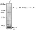
| Post Translational Modifications | Serine phosphorylation of IRS1 is a mechanism for insulin resistance. Ser-307, Ser-312, Ser-315, and Ser-323 phosphorylations inhibit insulin action through disruption of IRS1 interaction with the insulin receptor INSR. Phosphorylation of Tyr-896 is required for GRB2-binding. Phosphorylated by ALK. Phosphorylated at Ser-270, Ser-307, Ser-636 and Ser-1101 by RPS6KB1.phosphorylation induces accelerated degradation of IRS1. Phosphorylated on tyrosine residues in response to insulin. In skeletal muscles, dephosphorylated on Tyr-612 by TNS2 under anabolic conditions.dephosphorylation results in the proteasomal degradation of IRS1. Ubiquitinated by the Cul7-RING(FBXW8) complex in a mTOR-dependent manner, leading to its degradation: the Cul7-RING(FBXW8) complex recognizes and binds IRS1 previously phosphorylated by S6 kinase (RPS6KB1 or RPS6KB2). Ubiquitinated by TRAF4 through 'Lys-29' linkage.this ubiquitination regulates the interaction of IRS1 with IGFR and IRS1 tyrosine phosphorylation upon IGF1 stimulation. S-nitrosylation at by BLVRB inhibits its activity. |
| Function | Signaling adapter protein that participates in the signal transduction from two prominent receptor tyrosine kinases, insulin receptor/INSR and insulin-like growth factor I receptor/IGF1R. Plays therefore an important role in development, growth, glucose homeostasis as well as lipid metabolism. Upon phosphorylation by the insulin receptor, functions as a signaling scaffold that propagates insulin action through binding to SH2 domain-containing proteins including the p85 regulatory subunit of PI3K, NCK1, NCK2, GRB2 or SHP2. Recruitment of GRB2 leads to the activation of the guanine nucleotide exchange factor SOS1 which in turn triggers the Ras/Raf/MEK/MAPK signaling cascade. Activation of the PI3K/AKT pathway is responsible for most of insulin metabolic effects in the cell, and the Ras/Raf/MEK/MAPK is involved in the regulation of gene expression and in cooperation with the PI3K pathway regulates cell growth and differentiation. Acts a positive regulator of the Wnt/beta-catenin signaling pathway through suppression of DVL2 autophagy-mediated degradation leading to cell proliferation. |
| Protein Name | Insulin Receptor Substrate 1Irs-1 |
| Database Links | Reactome: R-HSA-109704Reactome: R-HSA-112399Reactome: R-HSA-112412Reactome: R-HSA-1257604Reactome: R-HSA-1266695Reactome: R-HSA-198203Reactome: R-HSA-201556Reactome: R-HSA-2219530Reactome: R-HSA-2428928Reactome: R-HSA-2586552Reactome: R-HSA-5673001Reactome: R-HSA-6811558Reactome: R-HSA-74713Reactome: R-HSA-74749Reactome: R-HSA-9603381Reactome: R-HSA-9725370Reactome: R-HSA-982772Reactome: R-HSA-9842663 |
| Cellular Localisation | CytoplasmNucleusNuclear Or Cytoplasmic Localization Of Irs1 Correlates With The Transition From Proliferation To Chondrogenic Differentiation |
| Alternative Antibody Names | Anti-Insulin Receptor Substrate 1 antibodyAnti-Irs-1 antibodyAnti-IRS1 antibody |
Information sourced from Uniprot.org


