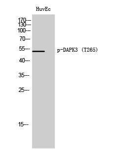
 Polyclonal Antibody STJ90708")
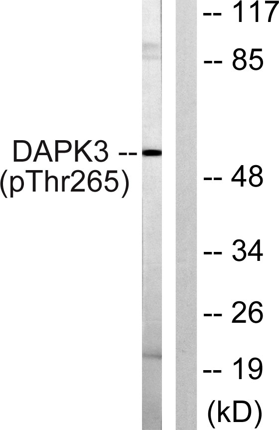
 Antibody. The lane on the right is STJ90708")
. 2, STJ90708")
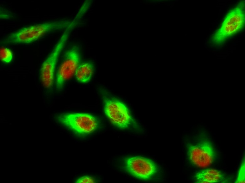
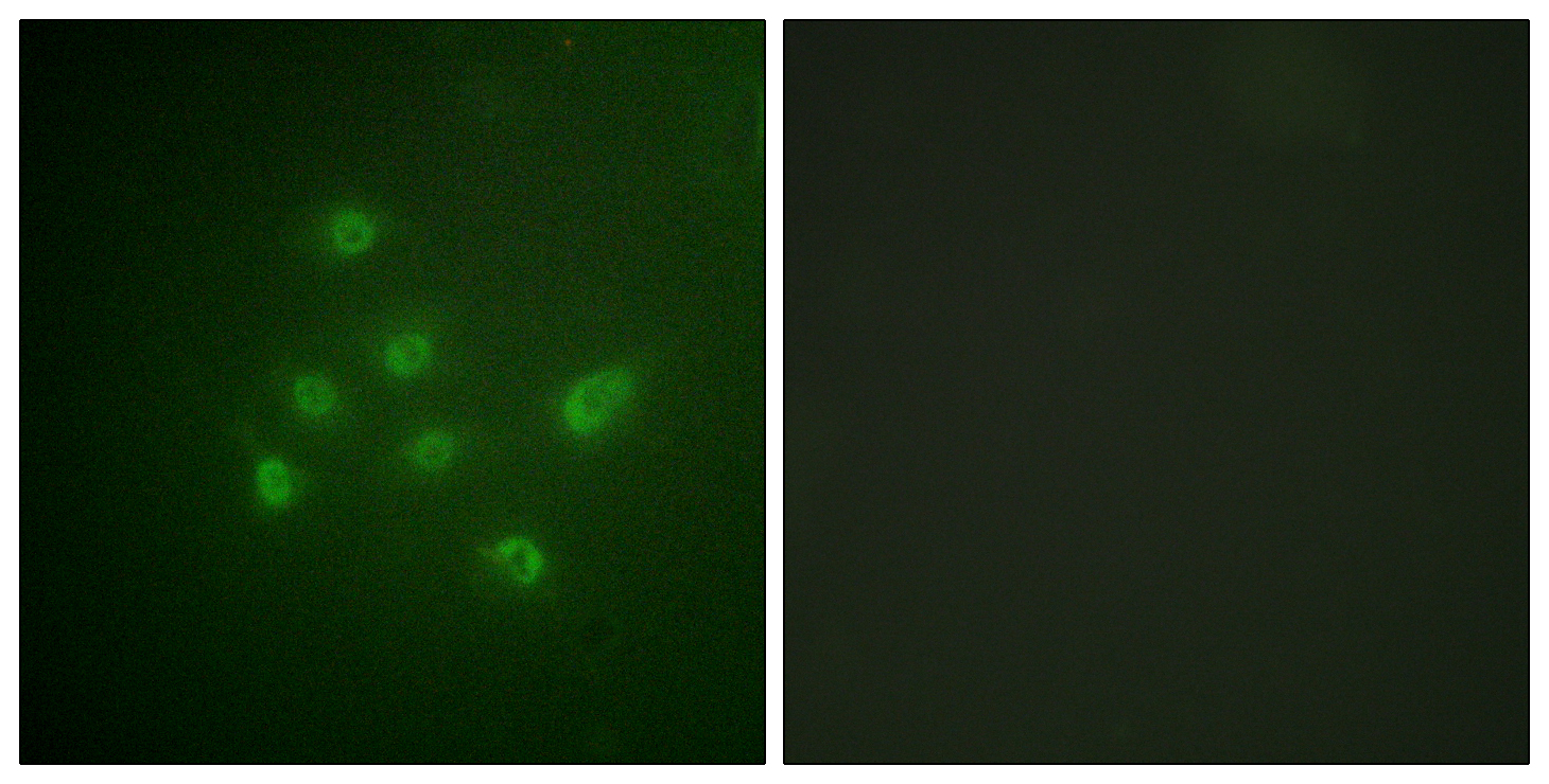
 Polyclonal Antibody (red) was diluted at 1:200 (4° STJ90708")
 Antibody. The picture on the right is blocked STJ90708")
 for Immunogen Phosphopeptide (Phospho-left) and Non-Phosphopeptide STJ90708")
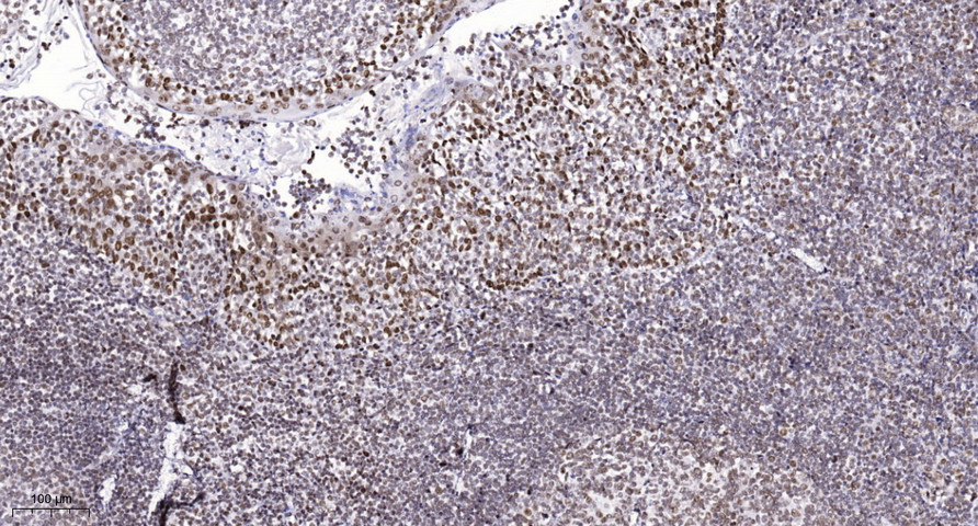
 Polyclonal Antibody STJ90708")
Anti-Phospho-DAPK3-Thr265 antibody (241-290 aa) (STJ90708)
SPECIFICATIONS
ClonalityPolyclonal
HostRabbit
ConjugationUnconjugated
IsotypeIgG
ImmunogenThe antiserum was produced against synthesized peptide derived from the human DAPK3 around the phosphorylation site of Thr265 at the amino acid range 241-290
General Information
| Short Description | Rabbit polyclonal anti-Phospho-Death-associated protein kinase 3-Thr265 (241-290 aa) for use in WB, IHC, IF and ELISA in Human, Mouse and Rat samples. Datasheet included with dilution recommendations, and related reagents. |
| Applications | WB/IHC/IF/ELISA |
| Host | Rabbit |
| Reactivity | Human/Mouse/Rat |
| Note | STRICTLY FOR FURTHER SCIENTIFIC RESEARCH USE ONLY (RUO). MUST NOT TO BE USED IN DIAGNOSTIC OR THERAPEUTIC APPLICATIONS. |
Product Properties
| Clonality | Polyclonal |
| Isotype | IgG |
| Conjugation | Unconjugated |
| Concentration | 1 mg/mL |
| Purification | The antibody was affinity-purified from rabbit antiserum by affinity-chromatography using epitope-specific immunogen. |
| Dilution Range | WB 1:500-1:2000IHC 1:100-1:300IF 1:200-1:1000ELISA 1:10000 |
| Formulation | Liquid in PBS containing 50% Glycerol, 0.5% BSA and 0.02% Sodium Azide. |
| Storage Instruction | Store at-20°C for up to 1 year from the date of receipt, and avoid repeat freeze-thaw cycles. |
Target Information
| Gene Symbol | DAPK3 |
| Gene ID | 1613 |
| Uniprot ID | DAPK3_HUMAN |
| Immunogen | The antiserum was produced against synthesized peptide derived from the human DAPK3 around the phosphorylation site of Thr265 at the amino acid range 241-290 |
| Immunogen Region | 241-290 aa |
| Specificity | Phospho-DAPK3 (T265) Polyclonal Antibody detects endogenous levels of DAPK3 protein only when phosphorylated at T265. |
Additional Info
| Post Translational Modifications | The phosphorylation status is critical for kinase activity, oligomerization and intracellular localization. Phosphorylation at Thr-180, Thr-225 and Thr-265 is essential for activity. The phosphorylated form is localized in the cytoplasm promoted by phosphorylation at Thr-299.nuclear translocation or retention is maximal when it is not phosphorylated. Phosphorylation increases the trimeric form, and its dephosphorylation favors a kinase-inactive monomeric form. Both isoform 1 and isoform 2 can undergo autophosphorylation. |
| Function | Serine/threonine kinase which is involved in the regulation of apoptosis, autophagy, transcription, translation and actin cytoskeleton reorganization. Involved in the regulation of smooth muscle contraction. Regulates both type I (caspase-dependent) apoptotic and type II (caspase-independent) autophagic cell deaths signal, depending on the cellular setting. Involved in regulation of starvation-induced autophagy. Regulates myosin phosphorylation in both smooth muscle and non-muscle cells. In smooth muscle, regulates myosin either directly by phosphorylating MYL12B and MYL9 or through inhibition of smooth muscle myosin phosphatase (SMPP1M) via phosphorylation of PPP1R12A.the inhibition of SMPP1M functions to enhance muscle responsiveness to Ca(2+) and promote a contractile state. Phosphorylates MYL12B in non-muscle cells leading to reorganization of actin cytoskeleton. Isoform 2 can phosphorylate myosin, PPP1R12A and MYL12B. Overexpression leads to condensation of actin stress fibers into thick bundles. Involved in actin filament focal adhesion dynamics. The function in both reorganization of actin cytoskeleton and focal adhesion dissolution is modulated by RhoD. Positively regulates canonical Wnt/beta-catenin signaling through interaction with NLK and TCF7L2. Phosphorylates RPL13A on 'Ser-77' upon interferon-gamma activation which is causing RPL13A release from the ribosome, RPL13A association with the GAIT complex and its subsequent involvement in transcript-selective translation inhibition. Enhances transcription from AR-responsive promoters in a hormone- and kinase-dependent manner. Involved in regulation of cell cycle progression and cell proliferation. May be a tumor suppressor. |
| Protein Name | Death-Associated Protein Kinase 3Dap Kinase 3Dap-Like KinaseDlkMypt1 KinaseZipper-Interacting Protein KinaseZip-Kinase |
| Database Links | Reactome: R-HSA-418889 |
| Cellular Localisation | NucleusPml BodyCytoplasmCytoskeletonMicrotubule Organizing CenterCentrosomeChromosomeCentromereSpindleMidbodyPredominantly Localizes To The Cytoplasm But Can Shuttle Between The Nucleus And CytoplasmCytoplasmic Localization Is Promoted By Phosphorylation At Thr-299 And Involves Rho/Rock SignalingIsoform 1: NucleusIsoform 2: Nucleus |
| Alternative Antibody Names | Anti-Death-Associated Protein Kinase 3 antibodyAnti-Dap Kinase 3 antibodyAnti-Dap-Like Kinase antibodyAnti-Dlk antibodyAnti-Mypt1 Kinase antibodyAnti-Zipper-Interacting Protein Kinase antibodyAnti-Zip-Kinase antibodyAnti-DAPK3 antibodyAnti-ZIPK antibody |
Information sourced from Uniprot.org