

Anti-PHH3 antibody (1-100aa) [ZR285] (STJ180477)
SPECIFICATIONS
ClonalityMonoclonal
HostRabbit
ConjugationUnconjugated
IsotypeIgG
ImmunogenSynthetic peptide corresponding to phosphohistone H3 residue within aa 1-100
General Information
| Short Description | Rabbit monoclonal PHH3 (1-100aa) antibody for use in IHC-P in human samples. Datasheet included with dilution recommendations, and related reagents. |
| Applications | IHC-P |
| Host | Rabbit |
| Reactivity | Human |
| Note | STRICTLY FOR FURTHER SCIENTIFIC RESEARCH USE ONLY (RUO). MUST NOT TO BE USED IN DIAGNOSTIC OR THERAPEUTIC APPLICATIONS. |
Product Properties
| Clonality | Monoclonal |
| Clone ID | ZR285 |
| Isotype | IgG |
| Conjugation | Unconjugated |
| Purification | Affinity purified |
| Dilution Range | 1:100-200 |
| Formulation | Tris-HCI buffer containing stabilizing protein (BSA) and <0.1% ProClin |
| Storage Instruction | Store at 2‐8°C for up to 24 months. Predilute: Ready to use, no reconstitution necessary. Concentrate: Use dilution range and appropriate lab‐standardized diluent. Stability after dilution: 7 days at 24°C, 3 months at 2‐8°C, 6months at ‐20°C. |
Target Information
| Gene Symbol | H3C15.H3C14.H3C13 |
| Gene ID | 126961/333932/653604 |
| Uniprot ID | H32_HUMAN |
| Immunogen | Synthetic peptide corresponding to phosphohistone H3 residue within aa 1-100 |
| Immunogen Region | 1-100aa |
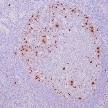
| Specificity | Positive control: Tonsil |
Additional Info
| Post Translational Modifications | Acetylation is generally linked to gene activation. Acetylation on Lys-10 (H3K9ac) impairs methylation at Arg-9 (H3R8me2s). Acetylation on Lys-19 (H3K18ac) and Lys-24 (H3K24ac) favors methylation at Arg-18 (H3R17me). Acetylation at Lys-123 (H3K122ac) by EP300/p300 plays a central role in chromatin structure: localizes at the surface of the histone octamer and stimulates transcription, possibly by promoting nucleosome instability. Citrullination at Arg-9 (H3R8ci) and/or Arg-18 (H3R17ci) by PADI4 impairs methylation and represses transcription. Asymmetric dimethylation at Arg-18 (H3R17me2a) by CARM1 is linked to gene activation. Symmetric dimethylation at Arg-9 (H3R8me2s) by PRMT5 is linked to gene repression. Asymmetric dimethylation at Arg-3 (H3R2me2a) by PRMT6 is linked to gene repression and is mutually exclusive with H3 Lys-5 methylation (H3K4me2 and H3K4me3). H3R2me2a is present at the 3' of genes regardless of their transcription state and is enriched on inactive promoters, while it is absent on active promoters. Methylation at Lys-5 (H3K4me), Lys-37 (H3K36me) and Lys-80 (H3K79me) are linked to gene activation. Methylation at Lys-5 (H3K4me) facilitates subsequent acetylation of H3 and H4. Methylation at Lys-80 (H3K79me) is associated with DNA double-strand break (DSB) responses and is a specific target for TP53BP1. Methylation at Lys-10 (H3K9me) and Lys-28 (H3K27me) are linked to gene repression. Methylation at Lys-10 (H3K9me) is a specific target for HP1 proteins (CBX1, CBX3 and CBX5) and prevents subsequent phosphorylation at Ser-11 (H3S10ph) and acetylation of H3 and H4. Methylation at Lys-5 (H3K4me) and Lys-80 (H3K79me) require preliminary monoubiquitination of H2B at 'Lys-120'. Methylation at Lys-10 (H3K9me) and Lys-28 (H3K27me) are enriched in inactive X chromosome chromatin. Monomethylation at Lys-57 (H3K56me1) by EHMT2/G9A in G1 phase promotes interaction with PCNA and is required for DNA replication. Phosphorylated at Thr-4 (H3T3ph) by VRK1. Phosphorylated at Thr-4 (H3T3ph) by HASPIN during prophase and dephosphorylated during anaphase. Phosphorylation at Ser-11 (H3S10ph) by AURKB is crucial for chromosome condensation and cell-cycle progression during mitosis and meiosis. In addition phosphorylation at Ser-11 (H3S10ph) by RPS6KA4 and RPS6KA5 is important during interphase because it enables the transcription of genes following external stimulation, like mitogens, stress, growth factors or UV irradiation and result in the activation of genes, such as c-fos and c-jun. Phosphorylation at Ser-11 (H3S10ph), which is linked to gene activation, prevents methylation at Lys-10 (H3K9me) but facilitates acetylation of H3 and H4. Phosphorylation at Ser-11 (H3S10ph) by AURKB mediates the dissociation of HP1 proteins (CBX1, CBX3 and CBX5) from heterochromatin. Phosphorylation at Ser-11 (H3S10ph) is also an essential regulatory mechanism for neoplastic cell transformation. Phosphorylated at Ser-29 (H3S28ph) by MAP3K20 isoform 1, RPS6KA5 or AURKB during mitosis or upon ultraviolet B irradiation. Phosphorylation at Thr-7 (H3T6ph) by PRKCB is a specific tag for epigenetic transcriptional activation that prevents demethylation of Lys-5 (H3K4me) by LSD1/KDM1A. At centromeres, specifically phosphorylated at Thr-12 (H3T11ph) from prophase to early anaphase, by DAPK3 and PKN1. Phosphorylation at Thr-12 (H3T11ph) by PKN1 or isoform M2 of PKM (PKM2) is a specific tag for epigenetic transcriptional activation that promotes demethylation of Lys-10 (H3K9me) by KDM4C/JMJD2C. Phosphorylation at Tyr-42 (H3Y41ph) by JAK2 promotes exclusion of CBX5 (HP1 alpha) from chromatin. Monoubiquitinated by RAG1 in lymphoid cells, monoubiquitination is required for V(D)J recombination. Ubiquitinated by the CUL4-DDB-RBX1 complex in response to ultraviolet irradiation. This may weaken the interaction between histones and DNA and facilitate DNA accessibility to repair proteins. Lysine deamination at Lys-5 (H3K4all) to form allysine is mediated by LOXL2. Allysine formation by LOXL2 only takes place on H3K4me3 and results in gene repression. Crotonylation (Kcr) is specifically present in male germ cells and marks testis-specific genes in post-meiotic cells, including X-linked genes that escape sex chromosome inactivation in haploid cells. Crotonylation marks active promoters and enhancers and confers resistance to transcriptional repressors. It is also associated with post-meiotically activated genes on autosomes. Butyrylation of histones marks active promoters and competes with histone acetylation. It is present during late spermatogenesis. Succinylation at Lys-80 (H3K79succ) by KAT2A takes place with a maximum frequency around the transcription start sites of genes. It gives a specific tag for epigenetic transcription activation. Desuccinylation at Lys-123 (H3K122succ) by SIRT7 in response to DNA damage promotes chromatin condensation and double-strand breaks (DSBs) repair. Serine ADP-ribosylation by PARP1 or PARP2 constitutes the primary form of ADP-ribosylation of proteins in response to DNA damage. Serine ADP-ribosylation at Ser-11 (H3S10ADPr) promotes recruitment of CHD1L. H3S10ADPr is mutually exclusive with phosphorylation at Ser-11 (H3S10ph) and impairs acetylation at Lys-10 (H3K9ac). Serotonylated by TGM2 at Gln-6 (H3Q5ser) during serotonergic neuron differentiation. H3Q5ser is associated with trimethylation of Lys-5 (H3K4me3) and enhances general transcription factor IID (TFIID) complex-binding to H3K4me3, thereby facilitating transcription. Dopaminylated by TGM2 at Gln-6 (H3Q5dop) in ventral tegmental area (VTA) neurons. H3Q5dop mediates neurotransmission-independent role of nuclear dopamine by regulating relapse-related transcriptional plasticity in the reward system. Lactylated in macrophages by EP300/P300 by using lactoyl-CoA directly derived from endogenous or exogenous lactate, leading to stimulates gene transcription. |
| Function | Core component of nucleosome. Nucleosomes wrap and compact DNA into chromatin, limiting DNA accessibility to the cellular machineries which require DNA as a template. Histones thereby play a central role in transcription regulation, DNA repair, DNA replication and chromosomal stability. DNA accessibility is regulated via a complex set of post-translational modifications of histones, also called histone code, and nucleosome remodeling. |
| Protein Name | Histone H3.2H3-Clustered Histone 13H3-Clustered Histone 14H3-Clustered Histone 15Histone H3/MHistone H3/O |
| Database Links | Reactome: R-HSA-1266695Reactome: R-HSA-1912408Reactome: R-HSA-201722Reactome: R-HSA-212300Reactome: R-HSA-2299718Reactome: R-HSA-2559580Reactome: R-HSA-2559582Reactome: R-HSA-3214815Reactome: R-HSA-3214841Reactome: R-HSA-3214842Reactome: R-HSA-3214847Reactome: R-HSA-3214858Reactome: R-HSA-3247509Reactome: R-HSA-427359Reactome: R-HSA-427389Reactome: R-HSA-427413Reactome: R-HSA-5250924Reactome: R-HSA-5334118Reactome: R-HSA-5578749Reactome: R-HSA-5617472Reactome: R-HSA-5625886Reactome: R-HSA-68616Reactome: R-HSA-73728Reactome: R-HSA-73772Reactome: R-HSA-8936459Reactome: R-HSA-8939236Reactome: R-HSA-9018519Reactome: R-HSA-912446Reactome: R-HSA-9609690Reactome: R-HSA-9610379Reactome: R-HSA-9616222Reactome: R-HSA-9710421Reactome: R-HSA-977225Reactome: R-HSA-9821002Reactome: R-HSA-983231Reactome: R-HSA-9841922Reactome: R-HSA-9843940Reactome: R-HSA-9843970Reactome: R-HSA-9845323 |
| Cellular Localisation | NucleusChromosome |
| Alternative Antibody Names | Anti-Histone H3.2 antibodyAnti-H3-Clustered Histone 13 antibodyAnti-H3-Clustered Histone 14 antibodyAnti-H3-Clustered Histone 15 antibodyAnti-Histone H3/M antibodyAnti-Histone H3/O antibodyAnti-H3C15 antibodyAnti-HIST2H3A.H3C14 antibodyAnti-H3F2 antibodyAnti-H3FM antibodyAnti-HIST2H3C.H3C13 antibodyAnti-HIST2H3D antibody |
Information sourced from Uniprot.org