

Anti-PD-1 antibody [DM177] (STJA0038180)
SPECIFICATIONS
ClonalityMonoclonal
HostRabbit
ConjugationUnconjugated
IsotypeIgG
ImmunogenRecombinant human PD-1 protein
General Information
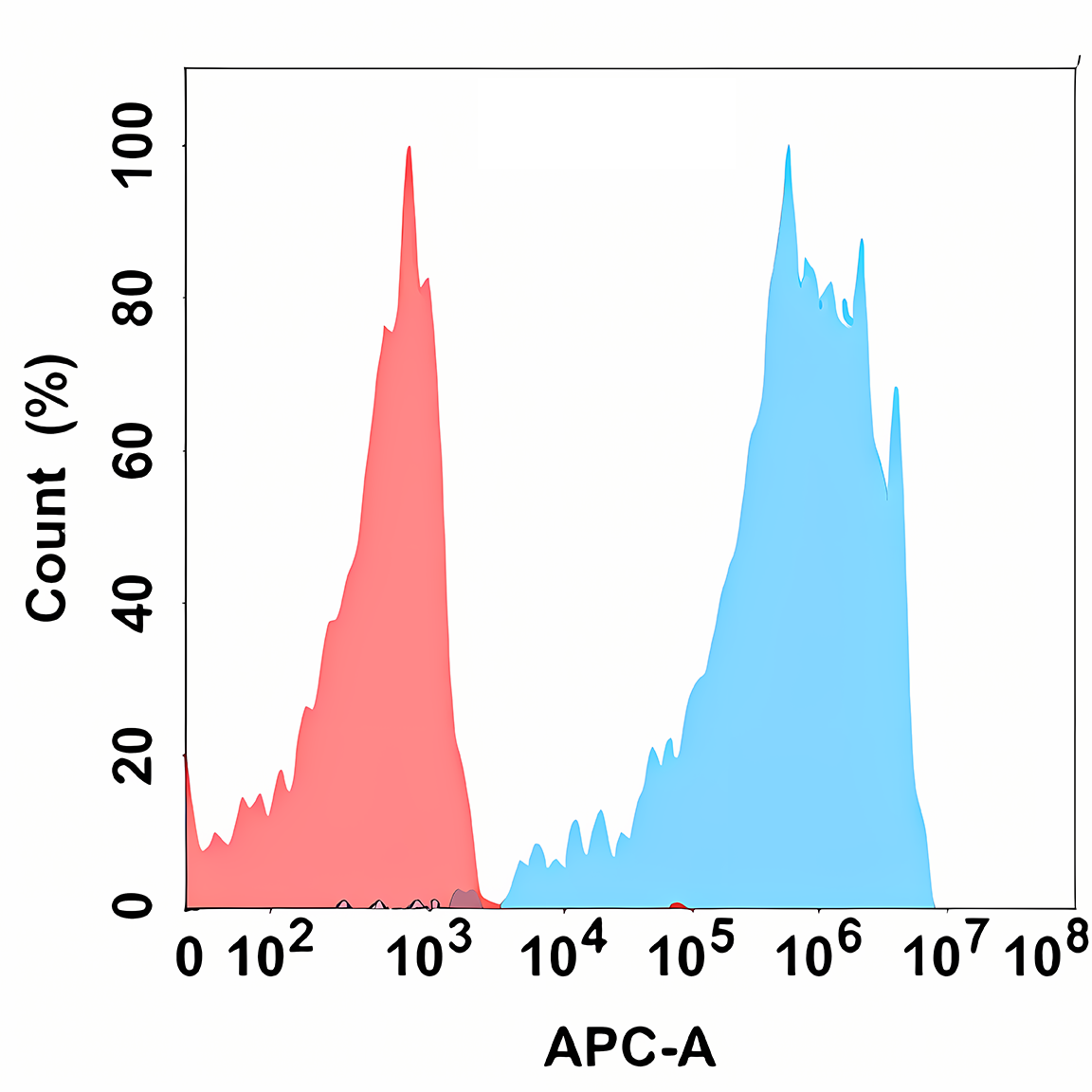
| Short Description | Rabbit monoclonal anti-PD-1 for use in FC and ELISA in Human samples. Datasheet included with dilution recommendations, and related reagents. |
| Applications | FC/ELISA |
| Host | Rabbit |
| Reactivity | Human |
| Note | STRICTLY FOR FURTHER SCIENTIFIC RESEARCH USE ONLY (RUO). MUST NOT TO BE USED IN DIAGNOSTIC OR THERAPEUTIC APPLICATIONS. |
Product Properties
| Clonality | Monoclonal |
| Clone ID | DM177 |
| Isotype | IgG |
| Conjugation | Unconjugated |
| Purification | Affinity Chromatography |
| Dilution Range | FC 1:50-1:100ELISA 1:10000 |
| Formulation | Liquid in PBS containing 50% glycerol, 0.5% BSA and 0.02% sodium azide, pH 7.3. |
| Storage Instruction | Store at 4°C short term. Aliquot and store at-20°C long term. Avoid freeze/thaw cycles. |
Target Information
| Gene Symbol | PDCD1 |
| Gene ID | 5133 |
| Uniprot ID | PDCD1_HUMAN |
| Immunogen | Recombinant human PD-1 protein |
Additional Info
| Post Translational Modifications | Ubiquitinated at Lys-233 by the SCF(FBXO38) complex, leading to its proteasomal degradation. Ubiquitinated via 'Lys-48'-linked polyubiquitin chains. Deubiquitinated and thus stabilized by USP5. Tyrosine phosphorylated at Tyr-223 (within ITIM motif) and Tyr-248 (ITSM motif) upon ligand binding. Phosphorylation at Tyr-248 promotes the recruitment of the protein tyrosine phosphatase PTPN11/SHP-2 that mediates dephosphorylation of key TCR proximal signaling molecules, such as ZAP70, PRKCQ/PKCtheta and CD247/CD3zeta. Phosphorylation at Thr-234 promotes the recruitment of the deubiquitinase USP5. N-glycosylation at Asn-58 contains at least two N-acetylglucosamine units and one fucose. N-glycosylation does not affect binding to nivolumab drug. |
| Function | Inhibitory receptor on antigen activated T-cells that plays a critical role in induction and maintenance of immune tolerance to self. Delivers inhibitory signals upon binding to ligands CD274/PDCD1L1 and CD273/PDCD1LG2. Following T-cell receptor (TCR) engagement, PDCD1 associates with CD3-TCR in the immunological synapse and directly inhibits T-cell activation. Suppresses T-cell activation through the recruitment of PTPN11/SHP-2: following ligand-binding, PDCD1 is phosphorylated within the ITSM motif, leading to the recruitment of the protein tyrosine phosphatase PTPN11/SHP-2 that mediates dephosphorylation of key TCR proximal signaling molecules, such as ZAP70, PRKCQ/PKCtheta and CD247/CD3zeta. The PDCD1-mediated inhibitory pathway is exploited by tumors to attenuate anti-tumor immunity and escape destruction by the immune system, thereby facilitating tumor survival. The interaction with CD274/PDCD1L1 inhibits cytotoxic T lymphocytes (CTLs) effector function. The blockage of the PDCD1-mediated pathway results in the reversal of the exhausted T-cell phenotype and the normalization of the anti-tumor response, providing a rationale for cancer immunotherapy. |
| Protein Name | Programmed Cell Death Protein 1Protein Pd-1Hpd-1Cd Antigen Cd279 |
| Database Links | Reactome: R-HSA-389948Reactome: R-HSA-9679191 |
| Cellular Localisation | Cell MembraneSingle-Pass Type I Membrane Protein |
| Alternative Antibody Names | Anti-Programmed Cell Death Protein 1 antibodyAnti-Protein Pd-1 antibodyAnti-Hpd-1 antibodyAnti-Cd Antigen Cd279 antibodyAnti-PDCD1 antibodyAnti-PD1 antibody |
Information sourced from Uniprot.org