


. STJ94441")


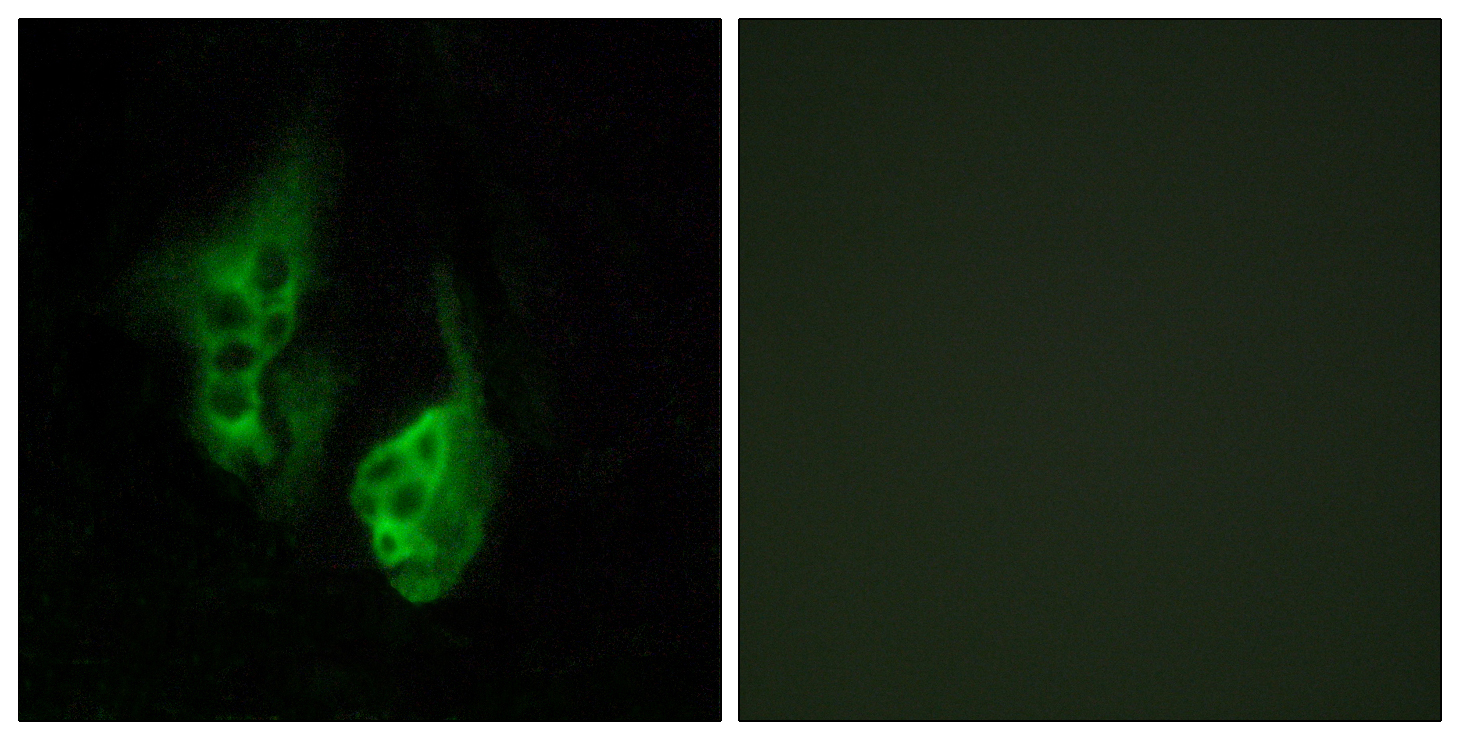
Anti-OPN5 antibody (251-300) (STJ94441)
SPECIFICATIONS
ClonalityPolyclonal
HostRabbit
ConjugationUnconjugated
IsotypeIgG
ImmunogenThe antiserum was produced against synthesized peptide derived from human OPN5. AA range:251-300
General Information
| Short Description | Rabbit polyclonal anti-Opsin-5 (251-300) for use in WB, ELISA and IHC in Human and Mouse samples. Datasheet included with dilution recommendations, and related reagents. |
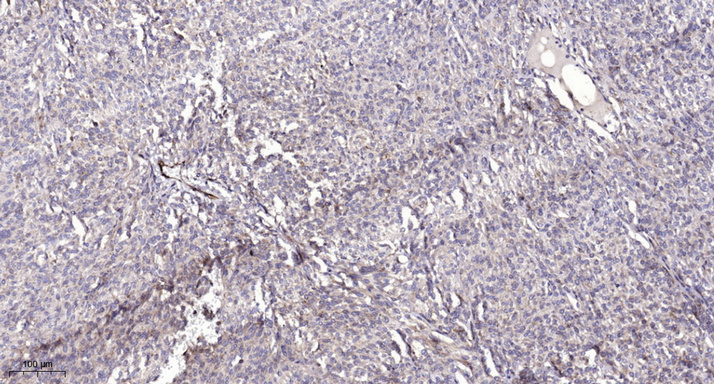
| Applications | WB/ELISA/IHC |
| Host | Rabbit |
| Reactivity | Human/Mouse |
| Note | STRICTLY FOR FURTHER SCIENTIFIC RESEARCH USE ONLY (RUO). MUST NOT TO BE USED IN DIAGNOSTIC OR THERAPEUTIC APPLICATIONS. |
Product Properties
| Clonality | Polyclonal |
| Isotype | IgG |
| Conjugation | Unconjugated |
| Concentration | 1 mg/mL |
| Purification | The antibody was affinity-purified from rabbit antiserum by affinity-chromatography using epitope-specific immunogen. |
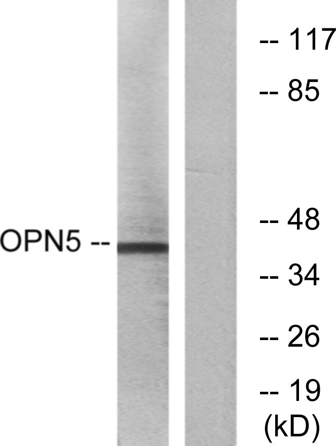
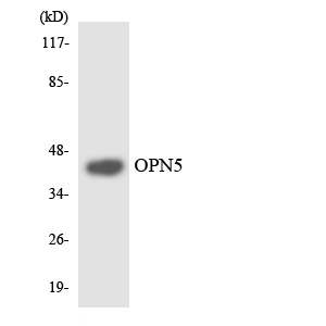
| Dilution Range | WB 1:500-2000IHC-p 1:50-300ELISA 2000-20000 |
| Formulation | Liquid in PBS containing 50% Glycerol, 0.5% BSA and 0.02% Sodium Azide. |
| Storage Instruction | Store at-20°C for up to 1 year from the date of receipt, and avoid repeat freeze-thaw cycles. |
Target Information
| Gene Symbol | OPN5 |
| Gene ID | 221391 |
| Uniprot ID | OPN5_HUMAN |
| Immunogen | The antiserum was produced against synthesized peptide derived from human OPN5. AA range:251-300 |
| Immunogen Region | 251-300 |
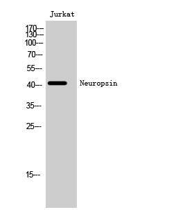
| Specificity | Neuropsin Polyclonal Antibody detects endogenous levels of Neuropsin protein. |
Additional Info
| Tissue Specificity | Detected in brain and retina and cell lines derived from neural retina. |
| Post Translational Modifications | It is uncertain whether Cys-315 or Cys-316 is palmitoylated. |
| Function | G-protein coupled receptor which selectively activates G(i) type G proteins via ultraviolet A (UVA) light-mediated activation in the retina. Preferentially binds the chromophore 11-cis retinal and is a bistable protein that displays emission peaks at 380 nm (UVA light) and 470 nm (blue light). Required for the light-response in the inner plexiform layer, and contributes to the regulation of the light-response in the nerve fiber layer, via phosphorylated DAT/SLC6A3 dopamine uptake. Involved in local corneal and retinal circadian rhythm photoentrainment via modulation of the UVA light-induced phase-shift of the retina clock. Acts as a circadian photoreceptor in the outer ear, via modulation of circadian clock-gene expression in response to violet light during the light-to-dark transition phase and night phase of the circadian cycle. Required in the retina to negatively regulate hyaloid vessel regression during postnatal development via light-dependent OPN5-SLC32A1-DRD2-VEGFR2 signaling. Involved in the light-dependent regulation of retina and vitreous compartment dopamine levels. |
| Protein Name | Opsin-5G-Protein Coupled Receptor 136G-Protein Coupled Receptor Pgr12NeuropsinTransmembrane Protein 13 |
| Database Links | Reactome: R-HSA-418594Reactome: R-HSA-419771 |
| Cellular Localisation | Cell MembraneMulti-Pass Membrane Protein |
| Alternative Antibody Names | Anti-Opsin-5 antibodyAnti-G-Protein Coupled Receptor 136 antibodyAnti-G-Protein Coupled Receptor Pgr12 antibodyAnti-Neuropsin antibodyAnti-Transmembrane Protein 13 antibodyAnti-OPN5 antibodyAnti-GPR136 antibodyAnti-PGR12 antibodyAnti-TMEM13 antibody |
Information sourced from Uniprot.org