

. 2, STJ92919")


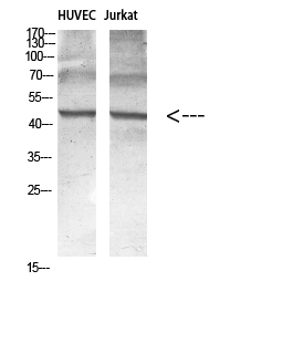
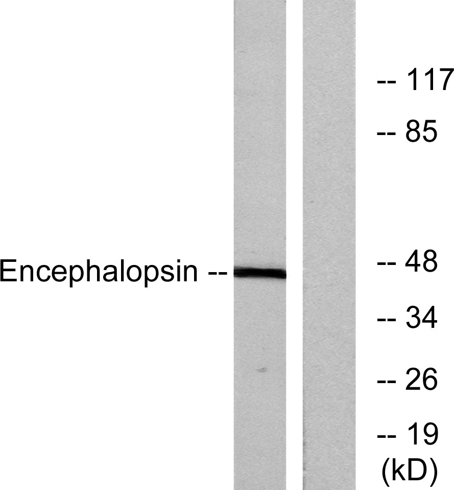
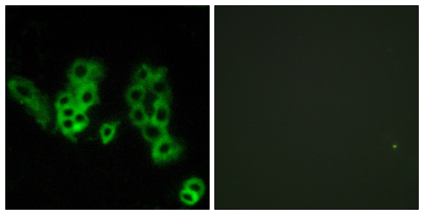
Anti-OPN3 antibody (161-210 aa) (STJ92919)
SPECIFICATIONS
ClonalityPolyclonal
HostRabbit
ConjugationUnconjugated
IsotypeIgG
ImmunogenThe antiserum was produced against synthesized peptide derived from the human Encephalopsin at the amino acid range 161-210
General Information
| Short Description | Rabbit polyclonal anti-Opsin-3 (161-210 aa) for use in WB, IHC, IF and ELISA in Human and Mouse samples. Datasheet included with dilution recommendations, and related reagents. |
| Applications | WB/IHC/IF/ELISA |
| Host | Rabbit |
| Reactivity | Human/Mouse |
| Note | STRICTLY FOR FURTHER SCIENTIFIC RESEARCH USE ONLY (RUO). MUST NOT TO BE USED IN DIAGNOSTIC OR THERAPEUTIC APPLICATIONS. |
Product Properties
| Clonality | Polyclonal |
| Isotype | IgG |
| Conjugation | Unconjugated |
| Concentration | 1 mg/mL |
| Purification | The antibody was affinity-purified from rabbit antiserum by affinity-chromatography using epitope-specific immunogen. |
| Dilution Range | WB 1:500-1:2000IHC 1:100-1:300IF 1:200-1:1000ELISA 1:5000 |
| Formulation | Liquid in PBS containing 50% Glycerol, 0.5% BSA and 0.02% Sodium Azide. |
| Storage Instruction | Store at-20°C for up to 1 year from the date of receipt, and avoid repeat freeze-thaw cycles. |
Target Information
| Gene Symbol | OPN3 |
| Gene ID | 23596 |
| Uniprot ID | OPN3_HUMAN |
| Immunogen | The antiserum was produced against synthesized peptide derived from the human Encephalopsin at the amino acid range 161-210 |
| Immunogen Region | 161-210 aa |
| Specificity | Encephalopsin Polyclonal Antibody detects endogenous levels of Encephalopsin protein. |
Additional Info
| Function | G-protein coupled receptor which selectively activates G proteins via ultraviolet A (UVA) light-mediated activation in the skin. Binds both 11-cis retinal and all-trans retinal. Regulates melanogenesis in melanocytes via inhibition of alpha-MSH-induced MC1R-mediated cAMP signaling, modulation of calcium flux, regulation of CAMK2 phosphorylation, and subsequently phosphorylation of CREB, p38, ERK and MITF in response to blue light. Plays a role in melanocyte survival through regulation of intracellular calcium levels and subsequent BCL2/RAF1 signaling. Additionally regulates apoptosis via cytochrome c release and subsequent activation of the caspase cascade. Required for TYR and DCT blue light-induced complex formation in melanocytes. Involved in keratinocyte differentiation in response to blue-light. Required for the UVA-mediated induction of calcium and mitogen-activated protein kinase signaling resulting in the expression of MMP1, MMP2, MMP3, MMP9 and TIMP1 in dermal fibroblasts. Plays a role in light-mediated glucose uptake, mitochondrial respiration and fatty acid metabolism in brown adipocyte tissues. May be involved in photorelaxation of airway smooth muscle cells, via blue-light dependent GPCR signaling pathways. |
| Protein Name | Opsin-3EncephalopsinPanopsin |
| Database Links | Reactome: R-HSA-418594Reactome: R-HSA-419771 |
| Cellular Localisation | Cell MembraneMulti-Pass Membrane ProteinCytoplasm |
| Alternative Antibody Names | Anti-Opsin-3 antibodyAnti-Encephalopsin antibodyAnti-Panopsin antibodyAnti-OPN3 antibodyAnti-ECPN antibody |
Information sourced from Uniprot.org