


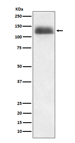
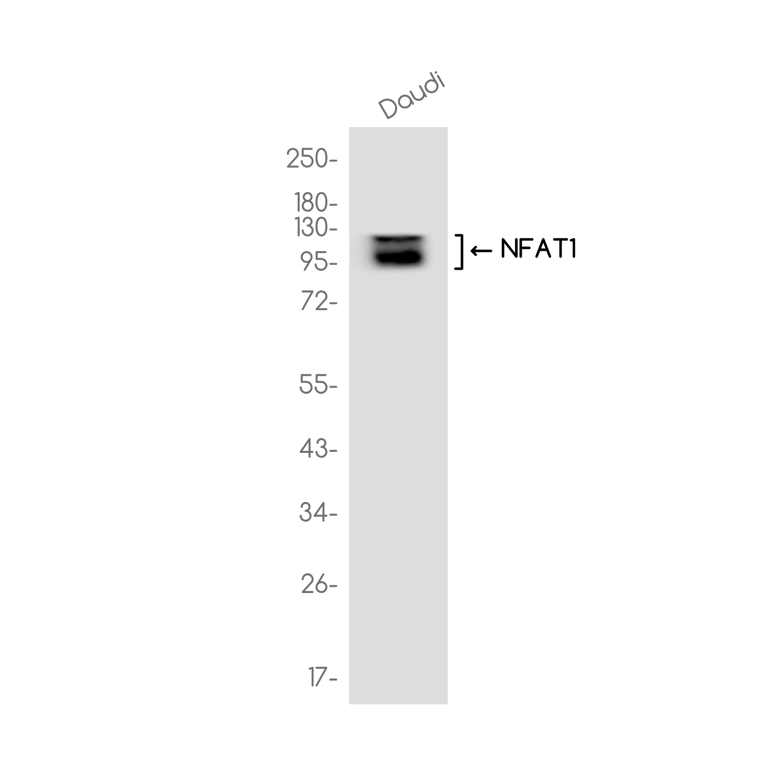
Anti-NFAT1 antibody [R04-1F7] (STJA0036360)
SPECIFICATIONS
ClonalityMonoclonal
HostRabbit
ConjugationUnconjugated
IsotypeIgG
ImmunogenA synthesized peptide derived from human NFAT1
General Information
| Short Description | Rabbit monoclonal anti-NFAT1 for use in WB in Human samples. Datasheet included with dilution recommendations, and related reagents. |
| Applications | WB |
| Host | Rabbit |
| Reactivity | Human |
| Note | STRICTLY FOR FURTHER SCIENTIFIC RESEARCH USE ONLY (RUO). MUST NOT TO BE USED IN DIAGNOSTIC OR THERAPEUTIC APPLICATIONS. |
Product Properties
| Clonality | Monoclonal |
| Clone ID | R04-1F7 |
| Isotype | IgG |
| Conjugation | Unconjugated |
| Purification | Affinity Chromatography |
| Dilution Range | WB 1:500-1:1000 |
| Formulation | Rabbit IgG in phosphate buffered saline, pH 7.4, 150mM NaCl, 0.02% sodium azide and 50% glycerol. |
| Storage Instruction | Store at 4°C short term. Aliquot and store at-20°C long term. Avoid freeze/thaw cycles. |
Target Information
| Gene Symbol | NFATC2 |
| Gene ID | 4773 |
| Uniprot ID | NFAC2_HUMAN |
| Immunogen | A synthesized peptide derived from human NFAT1 |
Additional Info
| Tissue Specificity | Expressed in thymus, spleen, heart, testis, brain, placenta, muscle and pancreas. Isoform 1 is highly expressed in the small intestine, heart, testis, prostate, thymus, placenta and thyroid. Isoform 3 is highly expressed in stomach, uterus, placenta, trachea and thyroid. |
| Post Translational Modifications | In resting cells, phosphorylated by NFATC-kinase on at least 18 sites in the 99-363 region. Upon cell stimulation, all these sites except Ser-243 are dephosphorylated by calcineurin. Dephosphorylation induces a conformational change that simultaneously exposes an NLS and masks an NES, which results in nuclear localization. Simultaneously, Ser-53 or Ser-56 is phosphorylated.which is required for full transcriptional activity. Ubiquitinated in endothelial cells by RNF213 downstream of the non-canonical Wnt signaling pathway, leading to its degradation by the proteasome. |
| Function | Plays a role in the inducible expression of cytokine genes in T-cells, especially in the induction of the IL-2, IL-3, IL-4, TNF-alpha or GM-CSF. Promotes invasive migration through the activation of GPC6 expression and WNT5A signaling pathway. Is involved in the negative regulation of chondrogenesis. Recruited by AKAP5 to ORAI1 pore-forming subunit of CRAC channels in Ca(2+) signaling microdomains where store-operated Ca(2+) influx is coupled to calmodulin and calcineurin signaling and activation of NFAT-dependent transcriptional responses. |
| Protein Name | Nuclear Factor Of Activated T-Cells - Cytoplasmic 2Nf-Atc2Nfatc2Nfat Pre-Existing SubunitNf-AtpT-Cell Transcription Factor Nfat1 |
| Database Links | Reactome: R-HSA-2025928Reactome: R-HSA-2871809Reactome: R-HSA-5607763Reactome: R-HSA-8877330 |
| Cellular Localisation | CytoplasmNucleusCytoplasmic For The Phosphorylated Form And Nuclear After Activation That Is Controlled By Calcineurin-Mediated DephosphorylationRapid Nuclear Exit Of Nfatc Is Thought To Be One Mechanism By Which Cells Distinguish Between Sustained And Transient Calcium SignalsThe Subcellular Localization Of Nfatc Plays A Key Role In The Regulation Of Gene Transcription |
| Alternative Antibody Names | Anti-Nuclear Factor Of Activated T-Cells - Cytoplasmic 2 antibodyAnti-Nf-Atc2 antibodyAnti-Nfatc2 antibodyAnti-Nfat Pre-Existing Subunit antibodyAnti-Nf-Atp antibodyAnti-T-Cell Transcription Factor Nfat1 antibodyAnti-NFATC2 antibodyAnti-NFAT1 antibodyAnti-NFATP antibody |
Information sourced from Uniprot.org