

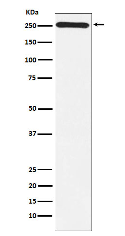
Anti-MYO7A antibody [R03-6V1] (STJA0036763)
SPECIFICATIONS
ClonalityMonoclonal
HostRabbit
ConjugationUnconjugated
IsotypeIgG
ImmunogenA synthesized peptide derived from human Myosin VIIa
General Information
| Short Description | Rabbit monoclonal anti-MYO7A for use in WB, ICC, IF and FC in Human, Mouse and Rat samples. Datasheet included with dilution recommendations, and related reagents. |
| Applications | WB/ICC/IF/FC |
| Host | Rabbit |
| Reactivity | Human/Mouse/Rat |
| Note | STRICTLY FOR FURTHER SCIENTIFIC RESEARCH USE ONLY (RUO). MUST NOT TO BE USED IN DIAGNOSTIC OR THERAPEUTIC APPLICATIONS. |
Product Properties
| Clonality | Monoclonal |
| Clone ID | R03-6V1 |
| Isotype | IgG |
| Conjugation | Unconjugated |
| Purification | Affinity Chromatography |
| Dilution Range | WB 1:500-1:1000IF 1:50-1:200FC 1:50-1:100 |
| Formulation | Rabbit IgG in phosphate buffered saline, pH 7.4, 150mM NaCl, 0.02% sodium azide and 50% glycerol. |
| Storage Instruction | Store at 4°C short term. Aliquot and store at-20°C long term. Avoid freeze/thaw cycles. |
Target Information
| Gene Symbol | MYO7A |
| Gene ID | 4647 |
| Uniprot ID | MYO7A_HUMAN |
| Immunogen | A synthesized peptide derived from human Myosin VIIa |
Additional Info
| Tissue Specificity | Expressed in the pigment epithelium and the photoreceptor cells of the retina. Also found in kidney, liver, testis, cochlea, lymphocytes. Not expressed in brain. |
| Function | Myosins are actin-based motor molecules with ATPase activity. Unconventional myosins serve in intracellular movements. Their highly divergent tails bind to membranous compartments, which are then moved relative to actin filaments. In the retina, plays an important role in the renewal of the outer photoreceptor disks. Plays an important role in the distribution and migration of retinal pigment epithelial (RPE) melanosomes and phagosomes, and in the regulation of opsin transport in retinal photoreceptors. In the inner ear, plays an important role in differentiation, morphogenesis and organization of cochlear hair cell bundles. Involved in hair-cell vesicle trafficking of aminoglycosides, which are known to induce ototoxicity. Motor protein that is a part of the functional network formed by USH1C, USH1G, CDH23 and MYO7A that mediates mechanotransduction in cochlear hair cells. Required for normal hearing. |
| Protein Name | Unconventional Myosin-Viia |
| Database Links | Reactome: R-HSA-2453902Reactome: R-HSA-9662360Reactome: R-HSA-9662361 |
| Cellular Localisation | CytoplasmCell CortexCytoskeletonSynapseIn The Photoreceptor CellsMainly Localized In The Inner And Base Of Outer Segments As Well As In The Synaptic Ending RegionIn Retinal Pigment Epithelial Cells Colocalizes With A Subset Of MelanosomesDisplays Predominant Localization To Stress Fiber-Like Structures And Some Localization To Cytoplasmic PunctaDetected At The Tip Of Cochlear Hair Cell StereociliaThe Complex Formed By Myo7aUsh1c And Ush1g Colocalizes With F-Actin |
| Alternative Antibody Names | Anti-Unconventional Myosin-Viia antibodyAnti-MYO7A antibodyAnti-USH1B antibody |
Information sourced from Uniprot.org