
. STJ97304")

Anti-MST1R antibody (31-80 aa) (STJ97304)
SPECIFICATIONS
ClonalityPolyclonal
HostRabbit
ConjugationUnconjugated
IsotypeIgG
ImmunogenThe antiserum was produced against synthesized peptide derived from the N-terminal region of human MST1R at the amino acid range 31-80
General Information
| Short Description | Rabbit polyclonal anti-Macrophage-stimulating protein receptor (31-80 aa) for use in WB and IHC in Human, Rat and Mouse samples. Datasheet included with dilution recommendations, and related reagents. |
| Applications | WB/IHC |
| Host | Rabbit |
| Reactivity | Human/Rat/Mouse |
| Note | STRICTLY FOR FURTHER SCIENTIFIC RESEARCH USE ONLY (RUO). MUST NOT TO BE USED IN DIAGNOSTIC OR THERAPEUTIC APPLICATIONS. |
Product Properties
| Clonality | Polyclonal |
| Isotype | IgG |
| Conjugation | Unconjugated |
| Concentration | 1 mg/mL |
| Purification | The antibody was affinity-purified from rabbit antiserum by affinity-chromatography using epitope-specific immunogen. |
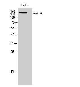
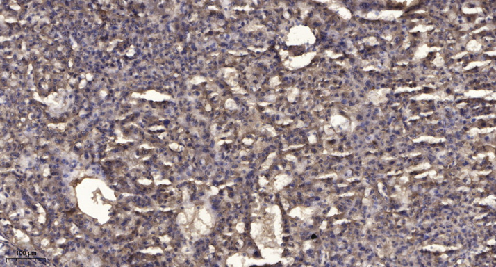
| Dilution Range | WB 1:500-2000IHC-P 1:50-300 |
| Formulation | Liquid in PBS containing 50% Glycerol, 0.5% BSA and 0.02% Sodium Azide. |
| Storage Instruction | Store at-20°C for up to 1 year from the date of receipt, and avoid repeat freeze-thaw cycles. |
Target Information
| Gene Symbol | MST1R |
| Gene ID | 4486 |
| Uniprot ID | RON_HUMAN |
| Immunogen | The antiserum was produced against synthesized peptide derived from the N-terminal region of human MST1R at the amino acid range 31-80 |
| Immunogen Region | 31-80 aa |
| Specificity | Ron Alpha Polyclonal Antibody detects endogenous levels of Ron Alpha protein. |
Additional Info
| Post Translational Modifications | Proteolytic processing yields the two subunits. Autophosphorylated in response to ligand binding on Tyr-1238 and Tyr-1239 in the kinase domain leading to further phosphorylation of Tyr-1353 and Tyr-1360 in the C-terminal multifunctional docking site. Ubiquitinated. Ubiquitination by CBL regulates the receptor stability and activity through proteasomal degradation. O-mannosylation of IPT/TIG domains on Thr or Ser residues by TMEM260 is required for protein maturation. O-mannosylated residues are composed of single mannose glycans that are not elongated or modified. |
| Function | Receptor tyrosine kinase that transduces signals from the extracellular matrix into the cytoplasm by binding to MST1 ligand. Regulates many physiological processes including cell survival, migration and differentiation. Ligand binding at the cell surface induces autophosphorylation of RON on its intracellular domain that provides docking sites for downstream signaling molecules. Following activation by ligand, interacts with the PI3-kinase subunit PIK3R1, PLCG1 or the adapter GAB1. Recruitment of these downstream effectors by RON leads to the activation of several signaling cascades including the RAS-ERK, PI3 kinase-AKT, or PLCgamma-PKC. RON signaling activates the wound healing response by promoting epithelial cell migration, proliferation as well as survival at the wound site. Also plays a role in the innate immune response by regulating the migration and phagocytic activity of macrophages. Alternatively, RON can also promote signals such as cell migration and proliferation in response to growth factors other than MST1 ligand. |
| Protein Name | Macrophage-Stimulating Protein ReceptorMsp ReceptorCdw136Protein-Tyrosine Kinase 8P185-RonCd Antigen Cd136 Cleaved Into - Macrophage-Stimulating Protein Receptor Alpha Chain - Macrophage-Stimulating Protein Receptor Beta Chain |
| Database Links | Reactome: R-HSA-8852405 |
| Cellular Localisation | MembraneSingle-Pass Type I Membrane Protein |
| Alternative Antibody Names | Anti-Macrophage-Stimulating Protein Receptor antibodyAnti-Msp Receptor antibodyAnti-Cdw136 antibodyAnti-Protein-Tyrosine Kinase 8 antibodyAnti-P185-Ron antibodyAnti-Cd Antigen Cd136 Cleaved Into - Macrophage-Stimulating Protein Receptor Alpha Chain - Macrophage-Stimulating Protein Receptor Beta Chain antibodyAnti-MST1R antibodyAnti-PTK8 antibodyAnti-RON antibody |
Information sourced from Uniprot.org