

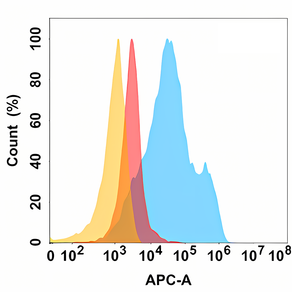
Anti-MICA antibody [DM157] (STJA0038173)
SPECIFICATIONS
ClonalityMonoclonal
HostRabbit
ConjugationUnconjugated
IsotypeIgG
ImmunogenRecombinant human MICA protein
General Information
| Short Description | Rabbit monoclonal anti-MICA for use in FC and ELISA in Human samples. Datasheet included with dilution recommendations, and related reagents. |
| Applications | FC/ELISA |
| Host | Rabbit |
| Reactivity | Human |
| Note | STRICTLY FOR FURTHER SCIENTIFIC RESEARCH USE ONLY (RUO). MUST NOT TO BE USED IN DIAGNOSTIC OR THERAPEUTIC APPLICATIONS. |
Product Properties
| Clonality | Monoclonal |
| Clone ID | DM157 |
| Isotype | IgG |
| Conjugation | Unconjugated |
| Purification | Affinity Chromatography |
| Dilution Range | FC 1:50-1:100ELISA 1:10000 |
| Formulation | Liquid in PBS containing 50% glycerol, 0.5% BSA and 0.02% sodium azide, pH 7.3. |
| Storage Instruction | Store at 4°C short term. Aliquot and store at-20°C long term. Avoid freeze/thaw cycles. |
Target Information
| Gene Symbol | MICA |
| Gene ID | 100507436 |
| Uniprot ID | MICA_HUMAN |
| Immunogen | Recombinant human MICA protein |
Additional Info
| Tissue Specificity | Widely expressed with the exception of the central nervous system where it is absent. Expressed predominantly in gastric epithelium and also in monocytes, keratinocytes, endothelial cells, fibroblasts and in the outer layer of Hassal's corpuscles within the medulla of normal thymus. In skin, expressed mainly in the keratin layers, basal cells, ducts and follicles. Also expressed in many, but not all, epithelial tumors of lung, breast, kidney, ovary, prostate and colon. In thyomas, overexpressed in cortical and medullar epithelial cells. Tumors expressing MICA display increased levels of gamma delta T-cells. |
| Post Translational Modifications | N-glycosylated. Glycosylation is not essential for interaction with KLRK1/NKG2D but enhances complex formation. Proteolytically cleaved and released from the cell surface of tumor cells which impairs KLRK1/NKG2D expression and T-cell activation. Palmitoylated on cysteine residues in the cytoplasmic tail leading to its association with membrane microdomains enriched in cholesterol. N-glycosylation is necessary for cell surface expression. (Microbial infection) Ubiquitinated by human herpesvirus 8 protein K5, leading to degradation. |
| Function | Widely expressed membrane-bound protein which acts as a ligand to stimulate an activating receptor KLRK1/NKG2D, expressed on the surface of essentially all human natural killer (NK), gammadelta T and CD8 alphabeta T-cells. Up-regulated in stressed conditions, such as viral and bacterial infections or DNA damage response, serves as signal of cellular stress, and engagement of KLRK1/NKG2D by MICA triggers NK-cells resulting in a range of immune effector functions, such as cytotoxicity and cytokine production. |
| Protein Name | Mhc Class I Polypeptide-Related Sequence AMic-A |
| Database Links | Reactome: R-HSA-198933 |
| Cellular Localisation | Cell MembraneSingle-Pass Type I Membrane ProteinCytoplasmExpressed On The Cell Surface In Gastric EpitheliumEndothelial Cells And Fibroblasts And In The Cytoplasm In Keratinocytes And MonocytesInfection With Human Adenovirus 5 Suppresses Cell Surface Expression Due To The Adenoviral E3-19k Protein Which Causes Retention In The Endoplasmic Reticulum |
| Alternative Antibody Names | Anti-Mhc Class I Polypeptide-Related Sequence A antibodyAnti-Mic-A antibodyAnti-MICA antibodyAnti-PERB11.1 antibody |
Information sourced from Uniprot.org