


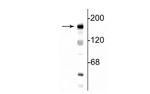
Anti-GRIN2B antibody (N-Term) (STJA0003718)
SPECIFICATIONS
ClonalityPolyclonal
HostRabbit
ConjugationUnconjugated
IsotypeIgG
ImmunogenSynthetic peptide corresponding to amino acid residues from the N-terminal region of the NR2B subunit conjugated to KLH.
General Information
| Short Description | Rabbit polyclonal anti-NMDAR2B (N-Term) for use in WB, IHC and ICC in Mouse, Rat, Bovine, Canine, Chicken, Human and Non-Human Primates samples. Datasheet included with dilution recommendations, and related reagents. |
| Applications | WB/IHC/ICC |
| Host | Rabbit |
| Reactivity | Mouse/Rat/Bovine/Canine/Chicken/Human/Non-Human Primates |
| Note | STRICTLY FOR FURTHER SCIENTIFIC RESEARCH USE ONLY (RUO). MUST NOT TO BE USED IN DIAGNOSTIC OR THERAPEUTIC APPLICATIONS. |
Product Properties
| Clonality | Polyclonal |
| Isotype | IgG |
| Conjugation | Unconjugated |
| Purification | This antibody was antigen affinity purified from pooled serum. |
| Dilution Range | WB 1:1000IHC 1:500ICC 1:400 |
| Formulation | 100 µl in 10 mM HEPES (pH 7.5) , 150 mM NaCl, 100 µg per ml BSA and 50% Glycerol. |
| Storage Instruction | Store at-20°C for up to 1 year from the date of receipt, and avoid repeat freeze-thaw cycles. |
Target Information
| Gene Symbol | Grin2b |
| Gene ID | 24410 |
| Uniprot ID | NMDE2_RAT |
| Immunogen | Synthetic peptide corresponding to amino acid residues from the N-terminal region of the NR2B subunit conjugated to KLH. |
| Immunogen Region | N-Term |
Additional Info
| Tissue Specificity | Expressed in the hippocampus including the dentate gyrus (at protein level). Detected in adult olfactory bulb, brain cortex, hippocampus, striatum, thalamus, superior colliculus, with much lower levels in inferior colliculus, midbrain and cerebellum. |
| Post Translational Modifications | Phosphorylated on tyrosine residues. Phosphorylation at Ser-1303 by DAPK1 enhances synaptic NMDA receptor channel activity. |
| Function | Component of N-methyl-D-aspartate (NMDA) receptors (NMDARs) that function as heterotetrameric, ligand-gated cation channels with high calcium permeability and voltage-dependent block by Mg(2+). Participates in synaptic plasticity for learning and memory formation by contributing to the long-term depression (LTD) of hippocampus membrane currents. Channel activation requires binding of the neurotransmitter L-glutamate to the GluN2 subunit, glycine or D-serine binding to the GluN1 subunit, plus membrane depolarization to eliminate channel inhibition by Mg(2+). NMDARs mediate simultaneously the potasium efflux and the influx of calcium and sodium. Each GluN2 subunit confers differential attributes to channel properties, including activation, deactivation and desensitization kinetics, pH sensitivity, Ca2(+) permeability, and binding to allosteric modulators. In concert with DAPK1 at extrasynaptic sites, acts as a central mediator for stroke damage. Its phosphorylation at Ser-1303 by DAPK1 enhances synaptic NMDA receptor channel activity inducing injurious Ca2+ influx through them, resulting in an irreversible neuronal death. |
| Protein Name | Glutamate Receptor Ionotropic - Nmda 2bGlun2bGlutamate Nmda Receptor Subunit Epsilon-2N-Methyl D-Aspartate Receptor Subtype 2bNmdar2bNr2b |
| Database Links | Reactome: R-RNO-3928662Reactome: -RNO-438066Reactome: -RNO-5673001Reactome: -RNO-8849932Reactome: -RNO-9609736 |
| Cellular Localisation | Cell MembraneMulti-Pass Membrane ProteinPostsynaptic Cell MembraneLate EndosomeLysosomeCytoplasmCytoskeletonCo-Localizes With The Motor Protein Kif17 Along Microtubules |
| Alternative Antibody Names | Anti-Glutamate Receptor Ionotropic - Nmda 2b antibodyAnti-Glun2b antibodyAnti-Glutamate Nmda Receptor Subunit Epsilon-2 antibodyAnti-N-Methyl D-Aspartate Receptor Subtype 2b antibodyAnti-Nmdar2b antibodyAnti-Nr2b antibodyAnti-Grin2b antibody |
Information sourced from Uniprot.org